 Movie
Movie Controller


+ Open data

- Basic information
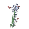
| Entry | Database: PDB / ID: 7du3 | ||||||
|---|---|---|---|---|---|---|---|
| Title | ThiL in complex with AMP-PNP | ||||||
 Components Components | Thiamine-monophosphate kinase | ||||||
 Keywords Keywords |  PROTEIN BINDING / Complex PROTEIN BINDING / Complex | ||||||
| Function / homology |  Function and homology informationthiamine-phosphate kinase / thiamine-phosphate kinase activity / thiamine diphosphate biosynthetic process / thiamine biosynthetic process / magnesium ion binding / ATP binding Function and homology informationthiamine-phosphate kinase / thiamine-phosphate kinase activity / thiamine diphosphate biosynthetic process / thiamine biosynthetic process / magnesium ion binding / ATP bindingSimilarity search - Function | ||||||
| Biological species |   Pseudomonas aeruginosa (bacteria) Pseudomonas aeruginosa (bacteria) | ||||||
| Method | X-RAY DIFFRACTION / SYNCHROTRON / MOLECULAR REPLACEMENT / Resolution: 2.54 Å | ||||||
 Authors Authors | Lin, J.Q. / Lescar, J. | ||||||
 Citation Citation | Journal: To Be Published Title: ThiL in complex with AMP-PNP Authors: Lin, J.Q. / Lescar, J. | ||||||
| History |
|
- Structure visualization
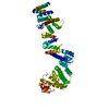
| Structure viewer | Molecule: MolmilJmol/JSmol |
|---|
- Downloads & links
-Download
| PDBx/mmCIF format | 7du3.cif.gz | 147.2 KB | Display | PDBx/mmCIF format |
|---|---|---|---|---|
| PDB format | pdb7du3.ent.gz | 103.3 KB | Display | PDB format |
| PDBx/mmJSON format | 7du3.json.gz | Tree view | PDBx/mmJSON format | |
| Others |  Other downloads Other downloads |
-Validation report
| Arichive directory | https://data.pdbj.org/pub/pdb/validation_reports/du/7du3ftp://data.pdbj.org/pub/pdb/validation_reports/du/7du3 | HTTPS FTP |
|---|
-Related structure data
| Related structure data | 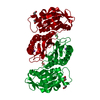 6xepS S: Starting model for refinement |
|---|---|
| Similar structure data |


-Links
 PDBj
PDBj


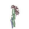
- Assembly
| Deposited unit | 
| ||||||||||||||||||||||||||||||
|---|---|---|---|---|---|---|---|---|---|---|---|---|---|---|---|---|---|---|---|---|---|---|---|---|---|---|---|---|---|---|---|
| 1 | 
| ||||||||||||||||||||||||||||||
| Unit cell |
| ||||||||||||||||||||||||||||||
| Components on special symmetry positions |
| ||||||||||||||||||||||||||||||
| Noncrystallographic symmetry (NCS) | NCS domain:
NCS domain segments:
NCS oper: (Code: givenMatrix: (0.00185795606976, -0.999395262971, 0.0347225625533), (-0.99802011519, -0.00403611155889, -0.062765910168), (0.0628680974341, -0.0345371995754, -0.997424074389)Vector: ...NCS oper: (Code: given Matrix: (0.00185795606976, -0.999395262971, 0.0347225625533), Vector : |
-Components
-Protein , 1 types, 2 molecules AB
| #1: Protein | Mass: 35495.383 Da / Num. of mol.: 2 Source method: isolated from a genetically manipulated source Source: (gene. exp.) Pseudomonas aeruginosa (bacteria)Gene: thiL, CAZ10_30480, DY930_26150, FDK04_05055, IPC116_27470, IPC1323_04495, IPC1509_03965, IPC582_16370, IPC620_29520, NCTC13621_06786 Production host: Escherichia coli (E. coli) / References: UniProt: A0A232BM78, thiamine-phosphate kinase |
|---|
-Non-polymers , 5 types, 171 molecules 


| #2: Chemical |  Mass: 506.196 Da / Num. of mol.: 2 / Source method: obtained synthetically / Formula: C10H17N6O12P3 / Feature type: SUBJECT OF INVESTIGATION / Comment: AMP-PNP, energy-carrying molecule analogue*YM Mass: 506.196 Da / Num. of mol.: 2 / Source method: obtained synthetically / Formula: C10H17N6O12P3 / Feature type: SUBJECT OF INVESTIGATION / Comment: AMP-PNP, energy-carrying molecule analogue*YM#3: Chemical | ChemComp-PG4 / | Polyethylene glycol Mass: 194.226 Da / Num. of mol.: 1 / Source method: obtained synthetically / Formula: C8H18O5 / Comment: precipitant*YM Mass: 194.226 Da / Num. of mol.: 1 / Source method: obtained synthetically / Formula: C8H18O5 / Comment: precipitant*YM#4: Chemical | ChemComp-EDO / | Ethylene glycol Mass: 62.068 Da / Num. of mol.: 1 / Source method: obtained synthetically / Formula: C2H6O2 Mass: 62.068 Da / Num. of mol.: 1 / Source method: obtained synthetically / Formula: C2H6O2#5: Chemical | ChemComp-MG /  Mass: 24.305 Da / Num. of mol.: 6 / Source method: obtained synthetically / Formula: Mg / Feature type: SUBJECT OF INVESTIGATION Mass: 24.305 Da / Num. of mol.: 6 / Source method: obtained synthetically / Formula: Mg / Feature type: SUBJECT OF INVESTIGATION#6: Water | ChemComp-HOH / | WaterMass: 18.015 Da / Num. of mol.: 161 / Source method: isolated from a natural source / Formula: H2O |
|---|
-Details
| Has ligand of interest | Y |
|---|
-Experimental details
-Experiment
| Experiment | Method: X-RAY DIFFRACTION / Number of used crystals: 1 |
|---|
- Sample preparation
| Crystal | Density Matthews: 3.05 Å3/Da / Density % sol: 59.64 % |
|---|---|
| Crystal grow | Temperature: 293.15 K / Method: vapor diffusion, sitting drop / pH: 8.5 Details: 0.02M DL-glutamic acid monohydrate, 0.02M DL-alanine, 0.02M glycine, 0.02M DL-lysine monohydrochloride, 0.02M DL-serine, 0.061M Tris, 0.039M Bicine, pH 8.5, 20% v/v PEG 500 MME, 10% w/v PEG 20000 |
-Data collection
| Diffraction | Mean temperature: 80 K / Serial crystal experiment: N |
|---|---|
| Diffraction source | Source: SYNCHROTRON / Site: Australian Synchrotron  / Beamline: MX2 / Wavelength: 0.9537 Å / Beamline: MX2 / Wavelength: 0.9537 Å |
| Detector | Type: ADSC QUANTUM 315r / Detector: CCD / Date: Nov 29, 2020 |
| Radiation | Protocol: SINGLE WAVELENGTH / Monochromatic (M) / Laue (L): M / Scattering type: x-ray |
| Radiation wavelength | Wavelength: 0.9537 Å / Relative weight: 1 |
| Reflection | Resolution: 2.5→48 Å / Num. obs: 28688 / % possible obs: 99.3 % / Redundancy: 13.7 % / Biso Wilson estimate: 51.94 Å2 / CC1/2: 0.998 / CC star: 1 / Rmerge(I) obs: 0.175 / Rpim(I) all: 0.04875 / Rrim(I) all: 0.1817 / Net I/σ(I): 16.75 |
| Reflection shell | Resolution: 2.543→2.634 Å / Redundancy: 12.8 % / Rmerge(I) obs: 1.367 / Mean I/σ(I) obs: 2.23 / Num. unique obs: 2670 / CC1/2: 0.732 / CC star: 0.919 / Rpim(I) all: 0.3884 / % possible all: 93.55 |
- Processing
| Software |
| |||||||||||||||||||||||||||||||||||||||||||||||||||||||||||||||||||||||||||||||||||||||||||||||||||||||||||||||||||||||||||||||||||||||||||||||||||
|---|---|---|---|---|---|---|---|---|---|---|---|---|---|---|---|---|---|---|---|---|---|---|---|---|---|---|---|---|---|---|---|---|---|---|---|---|---|---|---|---|---|---|---|---|---|---|---|---|---|---|---|---|---|---|---|---|---|---|---|---|---|---|---|---|---|---|---|---|---|---|---|---|---|---|---|---|---|---|---|---|---|---|---|---|---|---|---|---|---|---|---|---|---|---|---|---|---|---|---|---|---|---|---|---|---|---|---|---|---|---|---|---|---|---|---|---|---|---|---|---|---|---|---|---|---|---|---|---|---|---|---|---|---|---|---|---|---|---|---|---|---|---|---|---|---|---|---|---|
| Refinement | Method to determine structure: MOLECULAR REPLACEMENT Starting model: 6XEP Resolution: 2.54→43.59 Å / SU ML: 0.315 / Cross valid method: FREE R-VALUE / σ(F): 1.33 / Phase error: 22.5381 Stereochemistry target values: GeoStd + Monomer Library + CDL v1.2
| |||||||||||||||||||||||||||||||||||||||||||||||||||||||||||||||||||||||||||||||||||||||||||||||||||||||||||||||||||||||||||||||||||||||||||||||||||
| Solvent computation | Shrinkage radii: 0.9 Å / VDW probe radii: 1.11 Å / Solvent model: FLAT BULK SOLVENT MODEL | |||||||||||||||||||||||||||||||||||||||||||||||||||||||||||||||||||||||||||||||||||||||||||||||||||||||||||||||||||||||||||||||||||||||||||||||||||
| Displacement parameters | Biso mean: 54.97 Å2 | |||||||||||||||||||||||||||||||||||||||||||||||||||||||||||||||||||||||||||||||||||||||||||||||||||||||||||||||||||||||||||||||||||||||||||||||||||
| Refinement step | Cycle: LAST / Resolution: 2.54→43.59 Å
| |||||||||||||||||||||||||||||||||||||||||||||||||||||||||||||||||||||||||||||||||||||||||||||||||||||||||||||||||||||||||||||||||||||||||||||||||||
| Refine LS restraints |
| |||||||||||||||||||||||||||||||||||||||||||||||||||||||||||||||||||||||||||||||||||||||||||||||||||||||||||||||||||||||||||||||||||||||||||||||||||
| Refine LS restraints NCS | Type: Torsion NCS / Rms dev position: 0.710477003323 Å | |||||||||||||||||||||||||||||||||||||||||||||||||||||||||||||||||||||||||||||||||||||||||||||||||||||||||||||||||||||||||||||||||||||||||||||||||||
| LS refinement shell |
|
