 Movie
Movie Controller


+ Open data

- Basic information
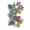
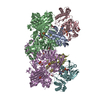
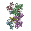
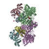
| Entry | Database: PDB / ID: 7dqx | ||||||
|---|---|---|---|---|---|---|---|
| Title | Crystal structure of xanthine dehydrogenase family protein | ||||||
 Components Components | (6-hydroxypseudooxynicotine dehydrogenase complex subunit ... ) x 4 ) x 4 | ||||||
 Keywords Keywords | OXIDOREDUCTASE / KDH | ||||||
| Function / homology |  Function and homology information6-hydroxypseudooxynicotine dehydrogenase / 6-hydroxypseudooxynicotine dehydrogenase activity / nicotine catabolic process / FAD binding / 2 iron, 2 sulfur cluster binding / metal ion binding Function and homology information6-hydroxypseudooxynicotine dehydrogenase / 6-hydroxypseudooxynicotine dehydrogenase activity / nicotine catabolic process / FAD binding / 2 iron, 2 sulfur cluster binding / metal ion bindingSimilarity search - Function | ||||||
| Biological species |  Paenarthrobacter nicotinovorans (bacteria) Paenarthrobacter nicotinovorans (bacteria) | ||||||
| Method | X-RAY DIFFRACTION / SYNCHROTRON / SAD / Resolution: 3.44 Å | ||||||
 Authors Authors | Lei, W. | ||||||
 Citation Citation | Journal: To Be Published Title: crystal structure of xanthine dehydrogenase family protein Authors: Lei, W. | ||||||
| History |
|
- Structure visualization
| Structure viewer | Molecule: MolmilJmol/JSmol |
|---|
- Downloads & links
-Download
| PDBx/mmCIF format | 7dqx.cif.gz | 900.8 KB | Display | PDBx/mmCIF format |
|---|---|---|---|---|
| PDB format | pdb7dqx.ent.gz | 737.2 KB | Display | PDB format |
| PDBx/mmJSON format | 7dqx.json.gz | Tree view | PDBx/mmJSON format | |
| Others |  Other downloads Other downloads |
-Validation report
| Arichive directory | https://data.pdbj.org/pub/pdb/validation_reports/dq/7dqxftp://data.pdbj.org/pub/pdb/validation_reports/dq/7dqx | HTTPS FTP |
|---|
-Related structure data
| Similar structure data |
|---|

-Links
 PDBj
PDBj


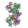
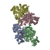
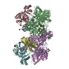
- Assembly
| Deposited unit | 
| ||||||||||||||||||||||||||||||||||||||||||||||||||||||||||||||||||||||||||||||||||||||||||||||||||||||
|---|---|---|---|---|---|---|---|---|---|---|---|---|---|---|---|---|---|---|---|---|---|---|---|---|---|---|---|---|---|---|---|---|---|---|---|---|---|---|---|---|---|---|---|---|---|---|---|---|---|---|---|---|---|---|---|---|---|---|---|---|---|---|---|---|---|---|---|---|---|---|---|---|---|---|---|---|---|---|---|---|---|---|---|---|---|---|---|---|---|---|---|---|---|---|---|---|---|---|---|---|---|---|---|
| 1 |
| ||||||||||||||||||||||||||||||||||||||||||||||||||||||||||||||||||||||||||||||||||||||||||||||||||||||
| Unit cell |
| ||||||||||||||||||||||||||||||||||||||||||||||||||||||||||||||||||||||||||||||||||||||||||||||||||||||
| Noncrystallographic symmetry (NCS) | NCS domain:
NCS domain segments:
NCS ensembles :
|
-Components
-6-hydroxypseudooxynicotine dehydrogenase complex subunit ... , 4 types, 6 molecules ADBCFE
| #1: Protein | / molybdopterin-binding subunit L / Ketone dehydrogenase large molybdopterin subunit Mass: 86474.555 Da / Num. of mol.: 2 Source method: isolated from a genetically manipulated source Source: (gene. exp.) Paenarthrobacter nicotinovorans (bacteria)Gene: kdhC, kdhL / Production host: Escherichia coli BL21(DE3) (bacteria)References: UniProt: Q933N0, 6-hydroxypseudooxynicotine dehydrogenase#2: Protein | | / molybdopterin-binding subunit M / Ketone dehydrogenase medium FAD subunitMass: 31469.957 Da / Num. of mol.: 1 Source method: isolated from a genetically manipulated source Source: (gene. exp.) Paenarthrobacter nicotinovorans (bacteria)Gene: kdhA, kdhM / Production host: Escherichia coli BL21(DE3) (bacteria)References: UniProt: O87681, 6-hydroxypseudooxynicotine dehydrogenase#3: Protein | / molybdopterin-binding subunit S / Ketone dehydrogenase small FeS subunitMass: 17660.904 Da / Num. of mol.: 2 Source method: isolated from a genetically manipulated source Source: (gene. exp.) Paenarthrobacter nicotinovorans (bacteria)Gene: kdhB, kdhS / Production host: Escherichia coli BL21(DE3) (bacteria)References: UniProt: O87682, 6-hydroxypseudooxynicotine dehydrogenase#4: Protein | | / molybdopterin-binding subunit M / Ketone dehydrogenase medium FAD subunitMass: 31469.957 Da / Num. of mol.: 1 Source method: isolated from a genetically manipulated source Source: (gene. exp.) Paenarthrobacter nicotinovorans (bacteria)Gene: kdhA, kdhM / Production host: Escherichia coli BL21(DE3) (bacteria)References: UniProt: O87681, 6-hydroxypseudooxynicotine dehydrogenase |
|---|
-Non-polymers , 4 types, 10 molecules 


| #5: Chemical |  Mass: 696.501 Da / Num. of mol.: 2 / Source method: obtained synthetically / Formula: C19H22N8O13P2S2 Mass: 696.501 Da / Num. of mol.: 2 / Source method: obtained synthetically / Formula: C19H22N8O13P2S2#6: Chemical |  Mass: 95.940 Da / Num. of mol.: 2 / Source method: obtained synthetically / Formula: Mo Mass: 95.940 Da / Num. of mol.: 2 / Source method: obtained synthetically / Formula: Mo#7: Chemical | Flavin adenine dinucleotide Mass: 785.550 Da / Num. of mol.: 2 / Source method: obtained synthetically / Formula: C27H33N9O15P2 / Comment: FAD*YM Mass: 785.550 Da / Num. of mol.: 2 / Source method: obtained synthetically / Formula: C27H33N9O15P2 / Comment: FAD*YM#8: Chemical | ChemComp-FES / Iron–sulfur cluster Mass: 175.820 Da / Num. of mol.: 4 / Source method: obtained synthetically / Formula: Fe2S2 Mass: 175.820 Da / Num. of mol.: 4 / Source method: obtained synthetically / Formula: Fe2S2 |
|---|
-Details
| Has ligand of interest | N |
|---|
-Experimental details
-Experiment
| Experiment | Method: X-RAY DIFFRACTION / Number of used crystals: 1 |
|---|
- Sample preparation
| Crystal | Density Matthews: 3.94 Å3/Da / Density % sol: 68.76 % |
|---|---|
| Crystal grow | Temperature: 293 K / Method: vapor diffusion, hanging drop / Details: PEG 4000 |
-Data collection
| Diffraction | Mean temperature: 80 K / Serial crystal experiment: N |
|---|---|
| Diffraction source | Source: SYNCHROTRON / Site: SSRF  / Beamline: BL17U1 / Wavelength: 0.979 Å / Beamline: BL17U1 / Wavelength: 0.979 Å |
| Detector | Type: Nonius Kappa CCD / Detector: CCD / Date: Dec 6, 2019 |
| Radiation | Protocol: SINGLE WAVELENGTH / Monochromatic (M) / Laue (L): M / Scattering type: x-ray |
| Radiation wavelength | Wavelength: 0.979 Å / Relative weight: 1 |
| Reflection | Resolution: 3.43→147.24 Å / Num. obs: 56814 / % possible obs: 96 % / Redundancy: 12.2 % / CC1/2: 0.87 / Net I/σ(I): 1.1 |
| Reflection shell | Resolution: 3.43→3.55 Å / Num. unique obs: 5553 / CC1/2: 0.512 |
- Processing
| Software |
| |||||||||||||||||||||||||||||||||||||||||||||||||||||||
|---|---|---|---|---|---|---|---|---|---|---|---|---|---|---|---|---|---|---|---|---|---|---|---|---|---|---|---|---|---|---|---|---|---|---|---|---|---|---|---|---|---|---|---|---|---|---|---|---|---|---|---|---|---|---|---|---|
| Refinement | Method to determine structure: SAD / Resolution: 3.44→147.24 Å / Cor.coef. Fo:Fc: 0.844 / Cor.coef. Fo:Fc free: 0.791 / SU B: 88.643 / SU ML: 0.625 / Cross valid method: THROUGHOUT / σ(F): 0 / ESU R Free: 0.709 / Stereochemistry target values: MAXIMUM LIKELIHOOD Details: HYDROGENS HAVE BEEN USED IF PRESENT IN THE INPUT U VALUES : REFINED INDIVIDUALLY
| |||||||||||||||||||||||||||||||||||||||||||||||||||||||
| Solvent computation | Ion probe radii: 0.8 Å / Shrinkage radii: 0.8 Å / VDW probe radii: 1.2 Å / Solvent model: MASK | |||||||||||||||||||||||||||||||||||||||||||||||||||||||
| Displacement parameters | Biso max: 153.74 Å2 / Biso mean: 67.87 Å2 / Biso min: 31.2 Å2
| |||||||||||||||||||||||||||||||||||||||||||||||||||||||
| Refinement step | Cycle: final / Resolution: 3.44→147.24 Å
| |||||||||||||||||||||||||||||||||||||||||||||||||||||||
| Refine LS restraints |
| |||||||||||||||||||||||||||||||||||||||||||||||||||||||
| Refine LS restraints NCS | Refine-ID: X-RAY DIFFRACTION / Type: interatomic distance / Weight position: 0.05
| |||||||||||||||||||||||||||||||||||||||||||||||||||||||
| LS refinement shell | Resolution: 3.443→3.532 Å / Rfactor Rfree error: 0 / Total num. of bins used: 20
|
