 Movie
Movie Controller

[English] 日本語
 Yorodumi
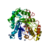
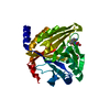
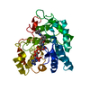
- PDB-7dp4: Crystal structure of wild type Brugia malayi thymidylate synthase... -
+ Open data

- Basic information
| Entry | Database: PDB / ID: 7dp4 | ||||||
|---|---|---|---|---|---|---|---|
| Title | Crystal structure of wild type Brugia malayi thymidylate synthase complexed with 2'-deoxyuridine monophosphate and methotrexate | ||||||
 Components Components | Thymidylate synthase | ||||||
 Keywords Keywords | TRANSFERASE / THYMIDYLATE SYNTHASE / NUCLEOTIDE SYNTHASE / METHYLTRANSFERASE | ||||||
| Function / homology |  Function and homology informationthymidylate synthase / thymidylate synthase activity / dTMP biosynthetic process / dTTP biosynthetic process / dihydrofolate reductase activity / methylation / mitochondrion / cytosol Function and homology informationthymidylate synthase / thymidylate synthase activity / dTMP biosynthetic process / dTTP biosynthetic process / dihydrofolate reductase activity / methylation / mitochondrion / cytosolSimilarity search - Function | ||||||
| Biological species |  Brugia malayi (agent of lymphatic filariasis) Brugia malayi (agent of lymphatic filariasis) | ||||||
| Method | X-RAY DIFFRACTION / SYNCHROTRON / MOLECULAR REPLACEMENT / Resolution: 1.5 Å | ||||||
 Authors Authors | Keyunratsami, K. / Nualnoi, T. / Wongkamchai, S. / Songsiriritthigul, C. / Chen, C.-J. / Canyuk, B. | ||||||
 Citation Citation | Journal: To Be Published Title: Crystallographic investigation and inhibition of Brugia malayi thymidylate synthase by a folate analog Authors: Keyunratsami, K. / Nualnoi, T. / Wongkamchai, S. / Songsiriritthigul, C. / Chen, C.-J. / Canyuk, B. | ||||||
| History |
|
- Structure visualization
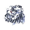
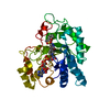
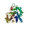
| Structure viewer | Molecule: MolmilJmol/JSmol |
|---|
- Downloads & links
-Download
| PDBx/mmCIF format | 7dp4.cif.gz | 86.1 KB | Display | PDBx/mmCIF format |
|---|---|---|---|---|
| PDB format | pdb7dp4.ent.gz | 60.1 KB | Display | PDB format |
| PDBx/mmJSON format | 7dp4.json.gz | Tree view | PDBx/mmJSON format | |
| Others |  Other downloads Other downloads |
-Validation report
| Arichive directory | https://data.pdbj.org/pub/pdb/validation_reports/dp/7dp4ftp://data.pdbj.org/pub/pdb/validation_reports/dp/7dp4 | HTTPS FTP |
|---|
-Related structure data
| Related structure data |  7dp5C  7dp6C 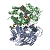 1hvyS S: Starting model for refinement C: citing same article ( |
|---|---|
| Similar structure data |


-Links
 PDBj
PDBj


- Assembly
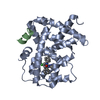
| Deposited unit | 
| ||||||||
|---|---|---|---|---|---|---|---|---|---|
| 1 |
| ||||||||
| Unit cell |
|
-Components
| #1: Protein | Mass: 34522.438 Da / Num. of mol.: 1 Source method: isolated from a genetically manipulated source Source: (gene. exp.) Brugia malayi (agent of lymphatic filariasis)Gene: Bma-tyms-1.1, BM_BM7277 / Plasmid: PQE-30 XA / Production host:  Escherichia coli (E. coli) / Strain (production host): SG13009 [PREP4] / References: UniProt: A0A4E9EZW9, thymidylate synthase Escherichia coli (E. coli) / Strain (production host): SG13009 [PREP4] / References: UniProt: A0A4E9EZW9, thymidylate synthase | ||||
|---|---|---|---|---|---|
| #2: Chemical | ChemComp-UMP / Deoxyuridine monophosphate  Mass: 308.182 Da / Num. of mol.: 1 / Source method: obtained synthetically / Formula: C9H13N2O8P Mass: 308.182 Da / Num. of mol.: 1 / Source method: obtained synthetically / Formula: C9H13N2O8P | ||||
| #3: Chemical | ChemComp-MTX / Methotrexate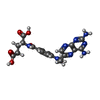  Mass: 454.439 Da / Num. of mol.: 1 / Source method: obtained synthetically / Formula: C20H22N8O5 / Comment: chemotherapy*YM Mass: 454.439 Da / Num. of mol.: 1 / Source method: obtained synthetically / Formula: C20H22N8O5 / Comment: chemotherapy*YM | ||||
| #4: Chemical | ChemComp-BME / 2-Mercaptoethanol  Mass: 78.133 Da / Num. of mol.: 5 / Source method: obtained synthetically / Formula: C2H6OS Mass: 78.133 Da / Num. of mol.: 5 / Source method: obtained synthetically / Formula: C2H6OS#5: Water | ChemComp-HOH / | Water Mass: 18.015 Da / Num. of mol.: 283 / Source method: isolated from a natural source / Formula: H2O Mass: 18.015 Da / Num. of mol.: 283 / Source method: isolated from a natural source / Formula: H2OHas ligand of interest | N | |
-Experimental details
-Experiment
| Experiment | Method: X-RAY DIFFRACTION / Number of used crystals: 1 |
|---|
- Sample preparation
| Crystal | Density Matthews: 2.92 Å3/Da / Density % sol: 57.9 % |
|---|---|
| Crystal grow | Temperature: 273 K / Method: vapor diffusion, hanging drop / pH: 7.5 / Details: 0.1 M HEPES, pH 7.5, 0.9 M sodium citrate |
-Data collection
| Diffraction | Mean temperature: 110 K / Serial crystal experiment: N |
|---|---|
| Diffraction source | Source: SYNCHROTRON / Site: NSRRC  / Beamline: TPS 05A / Wavelength: 1 Å / Beamline: TPS 05A / Wavelength: 1 Å |
| Detector | Type: MARMOSAIC 300 mm CCD / Detector: CCD / Date: Nov 12, 2013 / Details: DCM |
| Radiation | Monochromator: DCM / Protocol: SINGLE WAVELENGTH / Monochromatic (M) / Laue (L): M / Scattering type: x-ray |
| Radiation wavelength | Wavelength: 1 Å / Relative weight: 1 |
| Reflection | Resolution: 1.5→30 Å / Num. obs: 65291 / % possible obs: 99.7 % / Redundancy: 5.4 % / Rmerge(I) obs: 0.049 / Rpim(I) all: 0.033 / Rrim(I) all: 0.06 / Net I/σ(I): 26.06 |
| Reflection shell | Resolution: 1.5→1.55 Å / Redundancy: 5.4 % / Rmerge(I) obs: 0.491 / Mean I/σ(I) obs: 3.16 / Num. unique obs: 6480 / CC1/2: 0.718 / CC star: 0.914 / Rpim(I) all: 0.331 / Rrim(I) all: 0.597 / % possible all: 100 |
- Processing
| Software |
| |||||||||||||||||||||||||||||||||||||||||||||||||||||||||||||||||||||||||||||||||||||||||||||||||||||||||||||||||||||||||||||||||||||||||||||||||||||||||||
|---|---|---|---|---|---|---|---|---|---|---|---|---|---|---|---|---|---|---|---|---|---|---|---|---|---|---|---|---|---|---|---|---|---|---|---|---|---|---|---|---|---|---|---|---|---|---|---|---|---|---|---|---|---|---|---|---|---|---|---|---|---|---|---|---|---|---|---|---|---|---|---|---|---|---|---|---|---|---|---|---|---|---|---|---|---|---|---|---|---|---|---|---|---|---|---|---|---|---|---|---|---|---|---|---|---|---|---|---|---|---|---|---|---|---|---|---|---|---|---|---|---|---|---|---|---|---|---|---|---|---|---|---|---|---|---|---|---|---|---|---|---|---|---|---|---|---|---|---|---|---|---|---|---|---|---|---|
| Refinement | Method to determine structure: MOLECULAR REPLACEMENT Starting model: 1HVY Resolution: 1.5→25.855 Å / Cor.coef. Fo:Fc: 0.972 / Cor.coef. Fo:Fc free: 0.968 / SU B: 1.213 / SU ML: 0.045 / Cross valid method: FREE R-VALUE / ESU R: 0.062 / ESU R Free: 0.061 Details: Hydrogens have been added in their riding positions
| |||||||||||||||||||||||||||||||||||||||||||||||||||||||||||||||||||||||||||||||||||||||||||||||||||||||||||||||||||||||||||||||||||||||||||||||||||||||||||
| Solvent computation | Ion probe radii: 0.8 Å / Shrinkage radii: 0.8 Å / VDW probe radii: 1.2 Å / Solvent model: MASK BULK SOLVENT | |||||||||||||||||||||||||||||||||||||||||||||||||||||||||||||||||||||||||||||||||||||||||||||||||||||||||||||||||||||||||||||||||||||||||||||||||||||||||||
| Displacement parameters | Biso mean: 25.565 Å2
| |||||||||||||||||||||||||||||||||||||||||||||||||||||||||||||||||||||||||||||||||||||||||||||||||||||||||||||||||||||||||||||||||||||||||||||||||||||||||||
| Refinement step | Cycle: LAST / Resolution: 1.5→25.855 Å
| |||||||||||||||||||||||||||||||||||||||||||||||||||||||||||||||||||||||||||||||||||||||||||||||||||||||||||||||||||||||||||||||||||||||||||||||||||||||||||
| Refine LS restraints |
| |||||||||||||||||||||||||||||||||||||||||||||||||||||||||||||||||||||||||||||||||||||||||||||||||||||||||||||||||||||||||||||||||||||||||||||||||||||||||||
| LS refinement shell |
|
