 Movie
Movie Controller

[English] 日本語
 Yorodumi
- PDB-7dka: Crystal structure of DsbA-like protein DR2335 from Deinococcus ra... -
+ Open data

- Basic information
| Entry | Database: PDB / ID: 7dka | ||||||
|---|---|---|---|---|---|---|---|
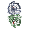
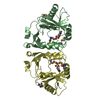
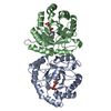
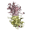
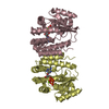
| Title | Crystal structure of DsbA-like protein DR2335 from Deinococcus radiodurans R1, C24S mutant protein | ||||||
 Components Components | DSBA domain-containing protein | ||||||
 Keywords Keywords |  OXIDOREDUCTASE / DsbA / thioredoxin fold / Thiol:disulfide interchange protein OXIDOREDUCTASE / DsbA / thioredoxin fold / Thiol:disulfide interchange protein | ||||||
| Function / homology | DSBA-like thioredoxin domain / DSBA-like thioredoxin domain / Thioredoxin-like superfamily / oxidoreductase activity / ACETATE ION / DI(HYDROXYETHYL)ETHER / DSBA domain-containing protein Function and homology information Function and homology information | ||||||
| Biological species |  Deinococcus radiodurans (radioresistant) Deinococcus radiodurans (radioresistant) | ||||||
| Method | X-RAY DIFFRACTION / SYNCHROTRON / MOLECULAR REPLACEMENT / Resolution: 1.56 Å | ||||||
 Authors Authors | Kim, M.-K. / Zhang, J. / Zhao, L. | ||||||
| Funding support |  Korea, Republic Of, 1items Korea, Republic Of, 1items
| ||||||
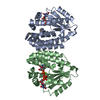
 Citation Citation | Journal: To Be Published Title: Crystal structure of DsbA-like protein DR2335 from Deinococcus radiodurans R1, native protein Authors: Kim, M.-K. / Zhang, J. / Zhao, L. | ||||||
| History |
|
- Structure visualization
| Structure viewer | Molecule: MolmilJmol/JSmol |
|---|
- Downloads & links
-Download
| PDBx/mmCIF format | 7dka.cif.gz | 113.3 KB | Display | PDBx/mmCIF format |
|---|---|---|---|---|
| PDB format | pdb7dka.ent.gz | 84.5 KB | Display | PDB format |
| PDBx/mmJSON format | 7dka.json.gz | Tree view | PDBx/mmJSON format | |
| Others |  Other downloads Other downloads |
-Validation report
| Arichive directory | https://data.pdbj.org/pub/pdb/validation_reports/dk/7dkaftp://data.pdbj.org/pub/pdb/validation_reports/dk/7dka | HTTPS FTP |
|---|
-Related structure data
| Related structure data |  7dk9SC S: Starting model for refinement C: citing same article ( |
|---|---|
| Similar structure data |


-Links
 PDBj
PDBj- Assembly
| Deposited unit | 
| ||||||||
|---|---|---|---|---|---|---|---|---|---|
| 1 | 
| ||||||||
| Unit cell |
|
-Components
| #1: Protein | Mass: 26238.105 Da / Num. of mol.: 2 / Mutation: C24S Source method: isolated from a genetically manipulated source Source: (gene. exp.) Deinococcus radiodurans (strain ATCC 13939 / DSM 20539 / JCM 16871 / LMG 4051 / NBRC 15346 / NCIMB 9279 / R1 / VKM B-1422) (radioresistant)Strain: ATCC 13939 / DSM 20539 / JCM 16871 / LMG 4051 / NBRC 15346 / NCIMB 9279 / R1 / VKM B-1422 Gene: DR_2335 / Production host: Escherichia coli (E. coli) / References: UniProt: Q9RRZ4#2: Chemical | ChemComp-ACT / Acetate  Mass: 59.044 Da / Num. of mol.: 12 / Source method: obtained synthetically / Formula: C2H3O2 Mass: 59.044 Da / Num. of mol.: 12 / Source method: obtained synthetically / Formula: C2H3O2#3: Chemical | Tris  Mass: 122.143 Da / Num. of mol.: 2 / Source method: obtained synthetically / Formula: C4H12NO3 / Comment: pH buffer*YM Mass: 122.143 Da / Num. of mol.: 2 / Source method: obtained synthetically / Formula: C4H12NO3 / Comment: pH buffer*YM#4: Chemical | Diethylene glycol  Mass: 106.120 Da / Num. of mol.: 3 / Source method: obtained synthetically / Formula: C4H10O3 Mass: 106.120 Da / Num. of mol.: 3 / Source method: obtained synthetically / Formula: C4H10O3#5: Water | ChemComp-HOH / | Water Mass: 18.015 Da / Num. of mol.: 526 / Source method: isolated from a natural source / Formula: H2O Mass: 18.015 Da / Num. of mol.: 526 / Source method: isolated from a natural source / Formula: H2OHas ligand of interest | N | |
|---|
-Experimental details
-Experiment
| Experiment | Method: X-RAY DIFFRACTION / Number of used crystals: 1 |
|---|
- Sample preparation
| Crystal | Density Matthews: 2.31 Å3/Da / Density % sol: 46.75 % |
|---|---|
| Crystal grow | Temperature: 295 K / Method: microbatch Details: Ammonium acetate, Sodium acetate trihydrate pH 4.6, 30% w/v PEG 4000 |
-Data collection
| Diffraction | Mean temperature: 100 K / Serial crystal experiment: N |
|---|---|
| Diffraction source | Source: SYNCHROTRON / Site: PAL/PLS / Beamline: 7A (6B, 6C1) / Wavelength: 0.97933 Å |
| Detector | Type: ADSC QUANTUM 270 / Detector: CCD / Date: May 15, 2019 |
| Radiation | Protocol: SINGLE WAVELENGTH / Monochromatic (M) / Laue (L): M / Scattering type: x-ray |
| Radiation wavelength | Wavelength: 0.97933 Å / Relative weight: 1 |
| Reflection | Resolution: 1.56→50 Å / Num. obs: 67292 / % possible obs: 99.9 % / Redundancy: 7.4 % / CC1/2: 0.986 / Rmerge(I) obs: 0.139 / Rpim(I) all: 0.055 / Rrim(I) all: 0.15 / Rsym value: 0.139 / Net I/σ(I): 37.635 |
| Reflection shell | Resolution: 1.56→1.59 Å / Rmerge(I) obs: 0.782 / Mean I/σ(I) obs: 4.845 / Num. unique obs: 3334 / CC1/2: 0.839 / Rpim(I) all: 0.31 / Rrim(I) all: 0.842 / Rsym value: 0.782 |
- Processing
| Software |
| |||||||||||||||||||||||||||||||||||||||||||||||||||||||||||||||||||||||||||||||||||||||||||||||||||||||||
|---|---|---|---|---|---|---|---|---|---|---|---|---|---|---|---|---|---|---|---|---|---|---|---|---|---|---|---|---|---|---|---|---|---|---|---|---|---|---|---|---|---|---|---|---|---|---|---|---|---|---|---|---|---|---|---|---|---|---|---|---|---|---|---|---|---|---|---|---|---|---|---|---|---|---|---|---|---|---|---|---|---|---|---|---|---|---|---|---|---|---|---|---|---|---|---|---|---|---|---|---|---|---|---|---|---|---|
| Refinement | Method to determine structure: MOLECULAR REPLACEMENT Starting model: 7DK9 Resolution: 1.56→29.86 Å / SU ML: 0.15 / Cross valid method: THROUGHOUT / σ(F): 1.34 / Phase error: 18.89 / Stereochemistry target values: ML
| |||||||||||||||||||||||||||||||||||||||||||||||||||||||||||||||||||||||||||||||||||||||||||||||||||||||||
| Solvent computation | Shrinkage radii: 0.9 Å / VDW probe radii: 1.11 Å / Solvent model: FLAT BULK SOLVENT MODEL | |||||||||||||||||||||||||||||||||||||||||||||||||||||||||||||||||||||||||||||||||||||||||||||||||||||||||
| Displacement parameters | Biso max: 83.4 Å2 / Biso mean: 19.9027 Å2 / Biso min: 6.16 Å2 | |||||||||||||||||||||||||||||||||||||||||||||||||||||||||||||||||||||||||||||||||||||||||||||||||||||||||
| Refinement step | Cycle: final / Resolution: 1.56→29.86 Å
| |||||||||||||||||||||||||||||||||||||||||||||||||||||||||||||||||||||||||||||||||||||||||||||||||||||||||
| LS refinement shell | Refine-ID: X-RAY DIFFRACTION / Rfactor Rfree error: 0 / Total num. of bins used: 14
|
