 Movie
Movie Controller

[English] 日本語
 Yorodumi
- PDB-7djs: Crystal structure of isopiperitenol dehydrogenase from Pseudomona... -
+ Open data

- Basic information
| Entry | Database: PDB / ID: 7djs | ||||||
|---|---|---|---|---|---|---|---|
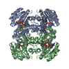
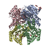
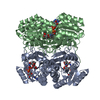
| Title | Crystal structure of isopiperitenol dehydrogenase from Pseudomonas aeruginosa complexed with NAD | ||||||
 Components Components | SDR family oxidoreductase | ||||||
 Keywords Keywords |  OXIDOREDUCTASE / complex / NAD OXIDOREDUCTASE / complex / NAD | ||||||
| Function / homology | Short-chain dehydrogenase/reductase, conserved site / Short-chain dehydrogenases/reductases family signature. / Short-chain dehydrogenase/reductase SDR / oxidoreductase activity / NAD(P)-binding domain superfamily / NICOTINAMIDE-ADENINE-DINUCLEOTIDE / SDR family oxidoreductase Function and homology information Function and homology information | ||||||
| Biological species |   Pseudomonas aeruginosa (bacteria) Pseudomonas aeruginosa (bacteria) | ||||||
| Method | X-RAY DIFFRACTION / SYNCHROTRON / MOLECULAR REPLACEMENT / Resolution: 1.7 Å | ||||||
 Authors Authors | Zhan, J.R. / Zheng, Y.C. | ||||||
 Citation Citation | Journal: Adv.Synth.Catal. / Year: 2021 Title: Discovery and Engineering of Bacterial (-)-Isopiperitenol Dehydrogenases to Enhance (-)-Menthol Precursor Biosynthesis. Authors: Zhan, J.R. / Shou, C. / Zheng, Y.C. / Chen, Q. / Pan, J. / Li, C.X. / Xu, J.H. | ||||||
| History |
|
- Structure visualization
| Structure viewer | Molecule: MolmilJmol/JSmol |
|---|
- Downloads & links
-Download
| PDBx/mmCIF format | 7djs.cif.gz | 206.5 KB | Display | PDBx/mmCIF format |
|---|---|---|---|---|
| PDB format | pdb7djs.ent.gz | 163 KB | Display | PDB format |
| PDBx/mmJSON format | 7djs.json.gz | Tree view | PDBx/mmJSON format | |
| Others |  Other downloads Other downloads |
-Validation report
| Arichive directory | https://data.pdbj.org/pub/pdb/validation_reports/dj/7djsftp://data.pdbj.org/pub/pdb/validation_reports/dj/7djs | HTTPS FTP |
|---|
-Related structure data
| Related structure data |  5x8hS S: Starting model for refinement |
|---|---|
| Similar structure data |


-Links
 PDBj
PDBj


- Assembly
| Deposited unit | 
| ||||||||
|---|---|---|---|---|---|---|---|---|---|
| 1 |
| ||||||||
| Unit cell |
|
-Components
| #1: Protein | Mass: 26812.752 Da / Num. of mol.: 4 Source method: isolated from a genetically manipulated source Source: (gene. exp.) Pseudomonas aeruginosa (bacteria)Gene: DY930_09865, F7O90_18305, FDK04_18370, IPC116_19850, IPC36_02985 Production host: Escherichia coli BL21(DE3) (bacteria) / References: UniProt: A0A554HE32#2: Chemical | ChemComp-NAD / Nicotinamide adenine dinucleotide  Mass: 663.425 Da / Num. of mol.: 4 / Source method: obtained synthetically / Formula: C21H27N7O14P2 / Comment: NAD*YM Mass: 663.425 Da / Num. of mol.: 4 / Source method: obtained synthetically / Formula: C21H27N7O14P2 / Comment: NAD*YM#3: Water | ChemComp-HOH / | Water Mass: 18.015 Da / Num. of mol.: 884 / Source method: isolated from a natural source / Formula: H2O Mass: 18.015 Da / Num. of mol.: 884 / Source method: isolated from a natural source / Formula: H2OHas ligand of interest | N | |
|---|
-Experimental details
-Experiment
| Experiment | Method: X-RAY DIFFRACTION / Number of used crystals: 1 |
|---|
- Sample preparation
| Crystal | Density Matthews: 2.12 Å3/Da / Density % sol: 42 % |
|---|---|
| Crystal grow | Temperature: 291 K / Method: vapor diffusion, sitting drop / pH: 6.25 Details: 100mM MES-imidazole pH 6.25, 30mM sodium nitrate, 30mM disodium hydrogen phosphate, 30mM ammonium sulfate, 20% v/v PEG 500MME, 10% w/v PEG 20000 PH range: 6.25-6.5 |
-Data collection
| Diffraction | Mean temperature: 100 K / Serial crystal experiment: N |
|---|---|
| Diffraction source | Source: SYNCHROTRON / Site: SSRF  / Beamline: BL19U1 / Wavelength: 0.9789 Å / Beamline: BL19U1 / Wavelength: 0.9789 Å |
| Detector | Type: DECTRIS PILATUS3 6M / Detector: PIXEL / Date: Oct 12, 2019 |
| Radiation | Protocol: SINGLE WAVELENGTH / Monochromatic (M) / Laue (L): M / Scattering type: x-ray |
| Radiation wavelength | Wavelength: 0.9789 Å / Relative weight: 1 |
| Reflection | Resolution: 1.7→50 Å / Num. obs: 100875 / % possible obs: 99.9 % / Redundancy: 13 % / CC1/2: 0.992 / Net I/σ(I): 27 |
| Reflection shell | Resolution: 1.7→1.73 Å / Num. unique obs: 5009 / CC1/2: 0.952 |
- Processing
| Software |
| |||||||||||||||||||||||||||||||||||||||||||||||||||||||||||||||||||||||||||||||||||||||||||||||||||||||||||||||||||||||||||||||||||||||||||||||||||||||||||||||||||||||||||||||||||||||||||||||||||||||||||||||||||||||||
|---|---|---|---|---|---|---|---|---|---|---|---|---|---|---|---|---|---|---|---|---|---|---|---|---|---|---|---|---|---|---|---|---|---|---|---|---|---|---|---|---|---|---|---|---|---|---|---|---|---|---|---|---|---|---|---|---|---|---|---|---|---|---|---|---|---|---|---|---|---|---|---|---|---|---|---|---|---|---|---|---|---|---|---|---|---|---|---|---|---|---|---|---|---|---|---|---|---|---|---|---|---|---|---|---|---|---|---|---|---|---|---|---|---|---|---|---|---|---|---|---|---|---|---|---|---|---|---|---|---|---|---|---|---|---|---|---|---|---|---|---|---|---|---|---|---|---|---|---|---|---|---|---|---|---|---|---|---|---|---|---|---|---|---|---|---|---|---|---|---|---|---|---|---|---|---|---|---|---|---|---|---|---|---|---|---|---|---|---|---|---|---|---|---|---|---|---|---|---|---|---|---|---|---|---|---|---|---|---|---|---|---|---|---|---|---|---|---|---|
| Refinement | Method to determine structure: MOLECULAR REPLACEMENT Starting model: 5X8H Resolution: 1.7→42.23 Å / SU ML: 0.16 / Cross valid method: THROUGHOUT / σ(F): 1.34 / Phase error: 17.18 / Stereochemistry target values: ML
| |||||||||||||||||||||||||||||||||||||||||||||||||||||||||||||||||||||||||||||||||||||||||||||||||||||||||||||||||||||||||||||||||||||||||||||||||||||||||||||||||||||||||||||||||||||||||||||||||||||||||||||||||||||||||
| Solvent computation | Shrinkage radii: 0.9 Å / VDW probe radii: 1.11 Å / Solvent model: FLAT BULK SOLVENT MODEL | |||||||||||||||||||||||||||||||||||||||||||||||||||||||||||||||||||||||||||||||||||||||||||||||||||||||||||||||||||||||||||||||||||||||||||||||||||||||||||||||||||||||||||||||||||||||||||||||||||||||||||||||||||||||||
| Displacement parameters | Biso max: 88.54 Å2 / Biso mean: 16.0461 Å2 / Biso min: 2.97 Å2 | |||||||||||||||||||||||||||||||||||||||||||||||||||||||||||||||||||||||||||||||||||||||||||||||||||||||||||||||||||||||||||||||||||||||||||||||||||||||||||||||||||||||||||||||||||||||||||||||||||||||||||||||||||||||||
| Refinement step | Cycle: final / Resolution: 1.7→42.23 Å
| |||||||||||||||||||||||||||||||||||||||||||||||||||||||||||||||||||||||||||||||||||||||||||||||||||||||||||||||||||||||||||||||||||||||||||||||||||||||||||||||||||||||||||||||||||||||||||||||||||||||||||||||||||||||||
| LS refinement shell | Refine-ID: X-RAY DIFFRACTION / Rfactor Rfree error: 0 / Total num. of bins used: 30
|
