 Movie
Movie Controller

[English] 日本語
 Yorodumi
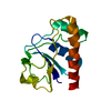
- PDB-7diu: Structure of PfGrx1 in the intermediate state with platinum and cesium -
+ Open data

- Basic information
| Entry | Database: PDB / ID: 7diu | ||||||
|---|---|---|---|---|---|---|---|
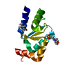
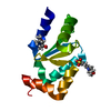
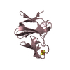
| Title | Structure of PfGrx1 in the intermediate state with platinum and cesium | ||||||
 Components Components | Glutaredoxin | ||||||
 Keywords Keywords | OXIDOREDUCTASE / REDOX ENZYME / TRX FOLD / GLUTATHIONE / Pt-SAD / Cs-SAD | ||||||
| Function / homology |  Function and homology informationprotein-disulfide reductase (glutathione) activity / Interconversion of nucleotide di- and triphosphates / glutathione disulfide oxidoreductase activity / antioxidant activity Function and homology informationprotein-disulfide reductase (glutathione) activity / Interconversion of nucleotide di- and triphosphates / glutathione disulfide oxidoreductase activity / antioxidant activitySimilarity search - Function | ||||||
| Biological species |  Plasmodium falciparum (malaria parasite P. falciparum) Plasmodium falciparum (malaria parasite P. falciparum) | ||||||
| Method | X-RAY DIFFRACTION / SAD / Resolution: 1.879 Å | ||||||
 Authors Authors | Manickam, Y. / Sharma, A. | ||||||
| Funding support |  India, 1items India, 1items
| ||||||
 Citation Citation | Journal: To Be Published Title: Interaction of metals with PfGrx1 Authors: Manickam, Y. / Sharma, A. | ||||||
| History |
|
- Structure visualization
| Structure viewer | Molecule: MolmilJmol/JSmol |
|---|
- Downloads & links
-Download
| PDBx/mmCIF format | 7diu.cif.gz | 57.6 KB | Display | PDBx/mmCIF format |
|---|---|---|---|---|
| PDB format | pdb7diu.ent.gz | 43.9 KB | Display | PDB format |
| PDBx/mmJSON format | 7diu.json.gz | Tree view | PDBx/mmJSON format | |
| Others |  Other downloads Other downloads |
-Validation report
| Arichive directory | https://data.pdbj.org/pub/pdb/validation_reports/di/7diuftp://data.pdbj.org/pub/pdb/validation_reports/di/7diu | HTTPS FTP |
|---|
-Related structure data
| Related structure data |  7dikC  7dilC  7dimC  7dinC  7dioC  7dipC  7diqC  7dirC 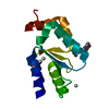 7disC  7ditC  7divC  7diwC  7dizC  7dj0C C: citing same article ( |
|---|---|
| Similar structure data |


-Links
 PDBj
PDBj
- Assembly
| Deposited unit | 
| ||||||||
|---|---|---|---|---|---|---|---|---|---|
| 1 |
| ||||||||
| Unit cell |
|
-Components
-Protein , 1 types, 1 molecules A
| #1: Protein | / Glutaredoxin 1 Mass: 12436.457 Da / Num. of mol.: 1 Source method: isolated from a genetically manipulated source Source: (gene. exp.) Plasmodium falciparum (isolate 3D7) (eukaryote)Strain: isolate 3D7 / Gene: PF3D7_0306300 / Plasmid: PQE30 / Production host:  Escherichia coli M15 (bacteria) Escherichia coli M15 (bacteria)References: UniProt: Q9NLB2, protein-disulfide reductase (glutathione) |
|---|
-Non-polymers , 5 types, 78 molecules 


| #2: Chemical | ChemComp-MPO / MOPS Mass: 209.263 Da / Num. of mol.: 1 / Source method: obtained synthetically / Formula: C7H15NO4S / Comment: pH buffer*YM Mass: 209.263 Da / Num. of mol.: 1 / Source method: obtained synthetically / Formula: C7H15NO4S / Comment: pH buffer*YM | ||||
|---|---|---|---|---|---|
| #3: Chemical | ChemComp-MPD / (2-Methyl-2,4-pentanediol Mass: 118.174 Da / Num. of mol.: 1 / Source method: obtained synthetically / Formula: C6H14O2 / Comment: precipitant*YM Mass: 118.174 Da / Num. of mol.: 1 / Source method: obtained synthetically / Formula: C6H14O2 / Comment: precipitant*YM | ||||
| #4: Chemical |  Mass: 132.905 Da / Num. of mol.: 3 / Source method: obtained synthetically / Formula: Cs / Feature type: SUBJECT OF INVESTIGATION Mass: 132.905 Da / Num. of mol.: 3 / Source method: obtained synthetically / Formula: Cs / Feature type: SUBJECT OF INVESTIGATION#5: Chemical | ChemComp-PT / |  Mass: 195.078 Da / Num. of mol.: 1 / Source method: obtained synthetically / Formula: Pt / Feature type: SUBJECT OF INVESTIGATION Mass: 195.078 Da / Num. of mol.: 1 / Source method: obtained synthetically / Formula: Pt / Feature type: SUBJECT OF INVESTIGATION#6: Water | ChemComp-HOH / | WaterMass: 18.015 Da / Num. of mol.: 72 / Source method: isolated from a natural source / Formula: H2O |
-Details
| Has ligand of interest | Y |
|---|
-Experimental details
-Experiment
| Experiment | Method: X-RAY DIFFRACTION / Number of used crystals: 1 |
|---|
- Sample preparation
| Crystal | Density Matthews: 2.27 Å3/Da / Density % sol: 45.72 % |
|---|---|
| Crystal grow | Temperature: 293 K / Method: vapor diffusion, hanging drop / pH: 7.5 Details: 12.5% w/v PEG1000, 12.5% w/v PEG3350, 12.5% v/v MPD, 0.02 M amino acids, 0.1 M MOPS/HEPES sodium |
-Data collection
| Diffraction | Mean temperature: 100 K / Serial crystal experiment: N | |||||||||||||||||||||||||||||||||||||||||||||||||||||||||||||||||||||||||||||||||||||||||||||||||||||||||||||||||||||||||||||||||||||||||||||||||||
|---|---|---|---|---|---|---|---|---|---|---|---|---|---|---|---|---|---|---|---|---|---|---|---|---|---|---|---|---|---|---|---|---|---|---|---|---|---|---|---|---|---|---|---|---|---|---|---|---|---|---|---|---|---|---|---|---|---|---|---|---|---|---|---|---|---|---|---|---|---|---|---|---|---|---|---|---|---|---|---|---|---|---|---|---|---|---|---|---|---|---|---|---|---|---|---|---|---|---|---|---|---|---|---|---|---|---|---|---|---|---|---|---|---|---|---|---|---|---|---|---|---|---|---|---|---|---|---|---|---|---|---|---|---|---|---|---|---|---|---|---|---|---|---|---|---|---|---|---|
| Diffraction source | Source: ROTATING ANODE / Type: RIGAKU MICROMAX-007 / Wavelength: 1.5418 Å | |||||||||||||||||||||||||||||||||||||||||||||||||||||||||||||||||||||||||||||||||||||||||||||||||||||||||||||||||||||||||||||||||||||||||||||||||||
| Detector | Type: MAR scanner 345 mm plate / Detector: IMAGE PLATE / Date: Jun 29, 2013 | |||||||||||||||||||||||||||||||||||||||||||||||||||||||||||||||||||||||||||||||||||||||||||||||||||||||||||||||||||||||||||||||||||||||||||||||||||
| Radiation | Protocol: SINGLE WAVELENGTH / Monochromatic (M) / Laue (L): M / Scattering type: x-ray | |||||||||||||||||||||||||||||||||||||||||||||||||||||||||||||||||||||||||||||||||||||||||||||||||||||||||||||||||||||||||||||||||||||||||||||||||||
| Radiation wavelength | Wavelength: 1.5418 Å / Relative weight: 1 | |||||||||||||||||||||||||||||||||||||||||||||||||||||||||||||||||||||||||||||||||||||||||||||||||||||||||||||||||||||||||||||||||||||||||||||||||||
| Reflection | Resolution: 1.879→50 Å / Num. obs: 17785 / % possible obs: 99.9 % / Redundancy: 20.7 % / Rmerge(I) obs: 0.066 / Χ2: 1.643 / Net I/σ(I): 15 | |||||||||||||||||||||||||||||||||||||||||||||||||||||||||||||||||||||||||||||||||||||||||||||||||||||||||||||||||||||||||||||||||||||||||||||||||||
| Reflection shell |
|
- Processing
| Software |
| ||||||||||||||||||||||||||||||||||||||||||||||||||||||||||||||||||||||||||||||||||||
|---|---|---|---|---|---|---|---|---|---|---|---|---|---|---|---|---|---|---|---|---|---|---|---|---|---|---|---|---|---|---|---|---|---|---|---|---|---|---|---|---|---|---|---|---|---|---|---|---|---|---|---|---|---|---|---|---|---|---|---|---|---|---|---|---|---|---|---|---|---|---|---|---|---|---|---|---|---|---|---|---|---|---|---|---|---|
| Refinement | Method to determine structure: SAD / Resolution: 1.879→27.574 Å / SU ML: 0.17 / Cross valid method: THROUGHOUT / σ(F): 1.35 / Phase error: 18.02 / Stereochemistry target values: ML
| ||||||||||||||||||||||||||||||||||||||||||||||||||||||||||||||||||||||||||||||||||||
| Solvent computation | Shrinkage radii: 0.9 Å / VDW probe radii: 1.11 Å / Solvent model: FLAT BULK SOLVENT MODEL | ||||||||||||||||||||||||||||||||||||||||||||||||||||||||||||||||||||||||||||||||||||
| Displacement parameters | Biso max: 86.13 Å2 / Biso mean: 31.3659 Å2 / Biso min: 16.34 Å2 | ||||||||||||||||||||||||||||||||||||||||||||||||||||||||||||||||||||||||||||||||||||
| Refinement step | Cycle: final / Resolution: 1.879→27.574 Å
| ||||||||||||||||||||||||||||||||||||||||||||||||||||||||||||||||||||||||||||||||||||
| LS refinement shell | Refine-ID: X-RAY DIFFRACTION / Rfactor Rfree error: 0
| ||||||||||||||||||||||||||||||||||||||||||||||||||||||||||||||||||||||||||||||||||||
| Refinement TLS params. | Method: refined / Origin x: -22.9593 Å / Origin y: 9.7755 Å / Origin z: -5.0076 Å
| ||||||||||||||||||||||||||||||||||||||||||||||||||||||||||||||||||||||||||||||||||||
| Refinement TLS group |
|
