 Movie
Movie Controller


+ Open data

- Basic information
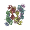
| Entry | Database: PDB / ID: 7dd9 | ||||||||||||
|---|---|---|---|---|---|---|---|---|---|---|---|---|---|
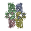
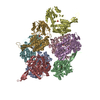
| Title | Cryo-EM structure of the Ams1 and Nbr1 complex | ||||||||||||
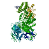
 Components Components | Alpha-mannosidase,ZZ-type zinc finger-containing protein P35G2.11c,Maltose/maltodextrin-binding periplasmic protein | ||||||||||||
 Keywords Keywords |  HYDROLASE / glycoside hydrolase / signaling protein / autophagy HYDROLASE / glycoside hydrolase / signaling protein / autophagy | ||||||||||||
| Function / homology |  Function and homology information Function and homology informationmannosyl-oligosaccharide 1,6-alpha-mannosidase activity / mannosyl-oligosaccharide 1,3-alpha-mannosidase activity / NVT complex / Lysosomal oligosaccharide catabolism / Pexophagy / alpha-mannosidase / fungal-type vacuole lumen / cytoplasm to vacuole targeting by the NVT pathway / alpha-mannosidase activity / oligosaccharide catabolic process ...mannosyl-oligosaccharide 1,6-alpha-mannosidase activity / mannosyl-oligosaccharide 1,3-alpha-mannosidase activity / NVT complex / Lysosomal oligosaccharide catabolism / Pexophagy / alpha-mannosidase / fungal-type vacuole lumen / cytoplasm to vacuole targeting by the NVT pathway / alpha-mannosidase activity / oligosaccharide catabolic process / mannose metabolic process / fungal-type vacuole membrane / cargo receptor activity / detection of maltose stimulus / maltose binding / maltose transport complex / maltose transport / maltodextrin transmembrane transport / carbohydrate transmembrane transporter activity / ATP-binding cassette (ABC) transporter complex, substrate-binding subunit-containing / carbohydrate transport / ATP-binding cassette (ABC) transporter complex / multivesicular body / cell chemotaxis / outer membrane-bounded periplasmic space / carbohydrate binding / periplasmic space / DNA damage response / Golgi apparatus / zinc ion binding / membrane / metal ion binding / cytosolSimilarity search - Function | ||||||||||||
| Biological species |  Schizosaccharomyces pombe (fission yeast) Schizosaccharomyces pombe (fission yeast) Escherichia coli (E. coli) Escherichia coli (E. coli) | ||||||||||||
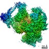
| Method | ELECTRON MICROSCOPY / single particle reconstruction / cryo EM / Resolution: 2.4 Å | ||||||||||||
 Authors Authors | Zhang, J. / Ye, K. | ||||||||||||
| Funding support |  China, 3items China, 3items
| ||||||||||||
 Citation Citation | Journal: EMBO J / Year: 2021 Title: Molecular and structural mechanisms of ZZ domain-mediated cargo selection by Nbr1. Authors: Ying-Ying Wang / Jianxiu Zhang / Xiao-Man Liu / Yulu Li / Jianhua Sui / Meng-Qiu Dong / Keqiong Ye / Li-Lin Du / Abstract: In selective autophagy, cargo selectivity is determined by autophagy receptors. However, it remains scarcely understood how autophagy receptors recognize specific protein cargos. In the fission yeast ...In selective autophagy, cargo selectivity is determined by autophagy receptors. However, it remains scarcely understood how autophagy receptors recognize specific protein cargos. In the fission yeast Schizosaccharomyces pombe, a selective autophagy pathway termed Nbr1-mediated vacuolar targeting (NVT) employs Nbr1, an autophagy receptor conserved across eukaryotes including humans, to target cytosolic hydrolases into the vacuole. Here, we identify two new NVT cargos, the mannosidase Ams1 and the aminopeptidase Ape4, that bind competitively to the first ZZ domain of Nbr1 (Nbr1-ZZ1). High-resolution cryo-EM analyses reveal how a single ZZ domain recognizes two distinct protein cargos. Nbr1-ZZ1 not only recognizes the N-termini of cargos via a conserved acidic pocket, similar to other characterized ZZ domains, but also engages additional parts of cargos in a cargo-specific manner. Our findings unveil a single-domain bispecific mechanism of autophagy cargo recognition, elucidate its underlying structural basis, and expand the understanding of ZZ domain-mediated protein-protein interactions. | ||||||||||||
| History |
|
- Structure visualization
| Movie |
Movie viewer |
|---|---|
| Structure viewer | Molecule: MolmilJmol/JSmol |
- Downloads & links
-Download
| PDBx/mmCIF format | 7dd9.cif.gz | 835.8 KB | Display | PDBx/mmCIF format |
|---|---|---|---|---|
| PDB format | pdb7dd9.ent.gz | 683.6 KB | Display | PDB format |
| PDBx/mmJSON format | 7dd9.json.gz | Tree view | PDBx/mmJSON format | |
| Others |  Other downloads Other downloads |
-Validation report
| Arichive directory | https://data.pdbj.org/pub/pdb/validation_reports/dd/7dd9ftp://data.pdbj.org/pub/pdb/validation_reports/dd/7dd9 | HTTPS FTP |
|---|
-Related structure data
| Related structure data |  30650MC  7ddeC M: map data used to model this data C: citing same article ( |
|---|---|
| Similar structure data |


-Links
 PDBj
PDBj


- Assembly
| Deposited unit | 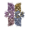
|
|---|---|
| 1 |
|
-Components
| #1: Protein | Mass: 178601.281 Da / Num. of mol.: 4 Source method: isolated from a genetically manipulated source Details: The fusion protein comprises of the full-length Ams1, a linker sequence (GFKKASSSDNKEQ), residues 53-180 of Nbr1, and maltose binding protein (MBP). Source: (gene. exp.) Schizosaccharomyces pombe (strain 972 / ATCC 24843) (yeast), (gene. exp.) Escherichia coli (strain K12) (bacteria)Gene: ams1, mns2, SPAC513.05, SPBP35G2.11c, malE, b4034, JW3994 Production host: Schizosaccharomyces pombe 972h- (yeast)References: UniProt: Q9UT61, UniProt: Q9P792, UniProt: P0AEX9, alpha-mannosidase#2: Chemical | ChemComp-ZN /   Mass: 65.409 Da / Num. of mol.: 12 / Source method: obtained synthetically / Formula: Zn / Feature type: SUBJECT OF INVESTIGATION Mass: 65.409 Da / Num. of mol.: 12 / Source method: obtained synthetically / Formula: Zn / Feature type: SUBJECT OF INVESTIGATION#3: Water | ChemComp-HOH / | Water Mass: 18.015 Da / Num. of mol.: 949 / Source method: isolated from a natural source / Formula: H2O Mass: 18.015 Da / Num. of mol.: 949 / Source method: isolated from a natural source / Formula: H2OHas ligand of interest | Y | |
|---|
-Experimental details
-Experiment
| Experiment | Method: ELECTRON MICROSCOPY |
|---|---|
| EM experiment | Aggregation state: PARTICLE / 3D reconstruction method: single particle reconstruction |
- Sample preparation
| Component | Name: Ams1 and Nbr1 fusion protein / Type: COMPLEX Details: The fusion protein comprises of the full-length Ams1, a linker sequence (GFKKASSSDNKEQ), residues 53-180 of Nbr1, and maltose binding protein (MBP). Entity ID: #1 / Source: RECOMBINANT | ||||||||||||||||||||
|---|---|---|---|---|---|---|---|---|---|---|---|---|---|---|---|---|---|---|---|---|---|
| Molecular weight | Value: 0.52 MDa / Experimental value: NO | ||||||||||||||||||||
| Source (natural) |
| ||||||||||||||||||||
| Source (recombinant) | Organism: Schizosaccharomyces pombe 972h- (yeast) | ||||||||||||||||||||
| Buffer solution | pH: 7.5 | ||||||||||||||||||||
| Buffer component |
| ||||||||||||||||||||
| Specimen | Conc.: 0.5 mg/ml / Embedding applied: NO / Shadowing applied: NO / Staining applied: NO / Vitrification applied: YES | ||||||||||||||||||||
| Specimen support | Grid material: GOLD / Grid mesh size: 300 divisions/in. / Grid type: Quantifoil R2/1 | ||||||||||||||||||||
| Vitrification | Instrument: FEI VITROBOT MARK IV / Cryogen name: ETHANE / Humidity: 100 % / Chamber temperature: 277 K / Details: blot for 5 seconds before plunging |
- Electron microscopy imaging
| Experimental equipment |  Model: Titan Krios / Image courtesy: FEI Company |
|---|---|
| Microscopy | Model: FEI TITAN KRIOS |
| Electron gun | Electron source: FIELD EMISSION GUN / Accelerating voltage: 300 kV / Illumination mode: FLOOD BEAM |
| Electron lens | Mode: BRIGHT FIELDBright-field microscopy / Nominal defocus max: 2200 nm / Nominal defocus min: 1600 nm / Cs: 2.7 mm |
| Specimen holder | Cryogen: NITROGEN / Specimen holder model: FEI TITAN KRIOS AUTOGRID HOLDER |
| Image recording | Average exposure time: 8.9 sec. / Electron dose: 60 e/Å2 / Detector mode: SUPER-RESOLUTION / Film or detector model: GATAN K2 SUMMIT (4k x 4k) / Num. of real images: 2430 |
| EM imaging optics | Energyfilter name: GIF Tridiem 4K / Energyfilter slit width: 20 eV |
| Image scans | Movie frames/image: 36 |
- Processing
| Software |
| ||||||||||||||||||||||||||||||||||||||||
|---|---|---|---|---|---|---|---|---|---|---|---|---|---|---|---|---|---|---|---|---|---|---|---|---|---|---|---|---|---|---|---|---|---|---|---|---|---|---|---|---|---|
| EM software |
| ||||||||||||||||||||||||||||||||||||||||
| CTF correction | Type: PHASE FLIPPING AND AMPLITUDE CORRECTION | ||||||||||||||||||||||||||||||||||||||||
| Symmetry | Point symmetry: D2 (2x2 fold dihedral) | ||||||||||||||||||||||||||||||||||||||||
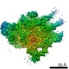
| 3D reconstruction | Resolution: 2.4 Å / Resolution method: FSC 0.143 CUT-OFF / Num. of particles: 296884 / Algorithm: FOURIER SPACE / Symmetry type: POINT | ||||||||||||||||||||||||||||||||||||||||
| Atomic model building | Protocol: AB INITIO MODEL / Space: REAL / Target criteria: Correlation coefficient | ||||||||||||||||||||||||||||||||||||||||
| Refinement | Cross valid method: NONE Stereochemistry target values: GeoStd + Monomer Library + CDL v1.2 | ||||||||||||||||||||||||||||||||||||||||
| Displacement parameters | Biso mean: 36.84 Å2 | ||||||||||||||||||||||||||||||||||||||||
| Refine LS restraints |
|
