 Movie
Movie Controller

[English] 日本語
 Yorodumi
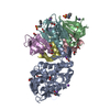
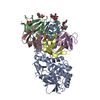
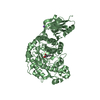
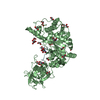
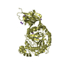
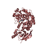
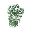
- PDB-7d6r: Crystal structure of the Stx2a complexed with MMA betaAla peptide -
+ Open data

- Basic information
| Entry | Database: PDB / ID: 7d6r | ||||||
|---|---|---|---|---|---|---|---|
| Title | Crystal structure of the Stx2a complexed with MMA betaAla peptide | ||||||
 Components Components |
| ||||||
 Keywords Keywords |  TOXIN / Shiga toxin TOXIN / Shiga toxin | ||||||
| Function / homology |  Function and homology information Function and homology informationhemolysis by symbiont of host erythrocytes / rRNA N-glycosylase / rRNA N-glycosylase activity / metabolic process / toxin activity / negative regulation of translation / extracellular regionSimilarity search - Function | ||||||
| Biological species |  Escherichia coli (E. coli) Escherichia coli (E. coli) | ||||||
| Method | X-RAY DIFFRACTION / SYNCHROTRON / MOLECULAR REPLACEMENT / Resolution: 1.6 Å | ||||||
 Authors Authors | Takahashi, M. / Tamada, M. / Hibino, M. / Senda, M. / Okuda, A. / Miyazawa, A. / Senda, T. / Nishikawa, K. | ||||||
| Funding support |  Japan, 1items Japan, 1items
| ||||||
 Citation Citation | Journal: Commun Biol / Year: 2021 Title: Identification of a peptide motif that potently inhibits two functionally distinct subunits of Shiga toxin. Authors: Watanabe-Takahashi, M. / Tamada, M. / Senda, M. / Hibino, M. / Shimizu, E. / Okuta, A. / Miyazawa, A. / Senda, T. / Nishikawa, K. | ||||||
| History |
|
- Structure visualization
| Structure viewer | Molecule: MolmilJmol/JSmol |
|---|
- Downloads & links
-Download
| PDBx/mmCIF format | 7d6r.cif.gz | 178.4 KB | Display | PDBx/mmCIF format |
|---|---|---|---|---|
| PDB format | pdb7d6r.ent.gz | 113.6 KB | Display | PDB format |
| PDBx/mmJSON format | 7d6r.json.gz | Tree view | PDBx/mmJSON format | |
| Others |  Other downloads Other downloads |
-Validation report
| Arichive directory | https://data.pdbj.org/pub/pdb/validation_reports/d6/7d6rftp://data.pdbj.org/pub/pdb/validation_reports/d6/7d6r | HTTPS FTP |
|---|
-Related structure data
| Related structure data | 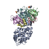 7d6qC  1r4pS S: Starting model for refinement C: citing same article ( |
|---|---|
| Similar structure data |
-Links
 PDBj
PDBj

- Assembly
| Deposited unit | 
| ||||||||||||
|---|---|---|---|---|---|---|---|---|---|---|---|---|---|
| 1 |
| ||||||||||||
| Unit cell |
|
-Components
| #1: Protein | / Shiga toxin 2 A subunit Mass: 33228.215 Da / Num. of mol.: 1 Source method: isolated from a genetically manipulated source Source: (gene. exp.) Escherichia coli (E. coli) / Gene: stx2a / Production host: Escherichia coli (E. coli) / References: UniProt: Q8XBV2, rRNA N-glycosylase | ||||||||||
|---|---|---|---|---|---|---|---|---|---|---|---|
| #2: Protein | Mass: 7824.590 Da / Num. of mol.: 5 Source method: isolated from a genetically manipulated source Source: (gene. exp.) Escherichia coli (E. coli) / Gene: stxII, stx2B, stx2B_2, stx2dB, stx2vB, stxB2, vtx2B / Production host: Escherichia coli (E. coli) / References: UniProt: Q7DJJ2#3: Protein/peptide | | Mass: 1251.619 Da / Num. of mol.: 1 Source method: isolated from a genetically manipulated source Source: (gene. exp.) Escherichia coli (E. coli) / Production host: Escherichia coli (E. coli)#4: Chemical | ChemComp-1PS / 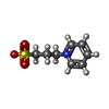  Mass: 201.243 Da / Num. of mol.: 4 / Source method: isolated from a natural source / Formula: C8H11NO3S Mass: 201.243 Da / Num. of mol.: 4 / Source method: isolated from a natural source / Formula: C8H11NO3S#5: Water | ChemComp-HOH / | Water Mass: 18.015 Da / Num. of mol.: 390 / Source method: isolated from a natural source / Formula: H2O Mass: 18.015 Da / Num. of mol.: 390 / Source method: isolated from a natural source / Formula: H2OHas ligand of interest | Y | Sequence details | Chain G has amidation of C-terminus. | |
-Experimental details
-Experiment
| Experiment | Method: X-RAY DIFFRACTION / Number of used crystals: 1 |
|---|
- Sample preparation
| Crystal | Density Matthews: 2.53 Å3/Da / Density % sol: 51.42 % |
|---|---|
| Crystal grow | Temperature: 293 K / Method: vapor diffusion, hanging drop Details: 4 M sodium formate, 100 mM MES pH 6.5, 50 mM 3-(1-Pyridinio)-1-propanesulfonate (PPS) |
-Data collection
| Diffraction | Mean temperature: 95 K / Serial crystal experiment: N |
|---|---|
| Diffraction source | Source: SYNCHROTRON / Site: Photon Factory / Beamline: BL-17A / Wavelength: 0.98 Å |
| Detector | Type: DECTRIS PILATUS3 S 6M / Detector: PIXEL / Date: Dec 11, 2015 |
| Radiation | Monochromator: Si 111 / Protocol: SINGLE WAVELENGTH / Monochromatic (M) / Laue (L): M / Scattering type: x-ray |
| Radiation wavelength | Wavelength: 0.98 Å / Relative weight: 1 |
| Reflection | Resolution: 1.6→73.25 Å / Num. obs: 97151 / % possible obs: 100 % / Redundancy: 9.8 % / Biso Wilson estimate: 17.82 Å2 / Rmerge(I) obs: 0.058 / Net I/σ(I): 21.48 |
| Reflection shell | Resolution: 1.6→1.69 Å / Rmerge(I) obs: 0.429 / Mean I/σ(I) obs: 6.37 / Num. unique obs: 14626 |
- Processing
| Software |
| |||||||||||||||||||||||||||||||||||||||||||||||||||||||||||||||||||||||||||||||||||||||||||||||||||||||||||||||||||||||||||||||||||||||||||||||||||||||||||||||||||||||||||||||||||||||||||||||||||||||||||||||||||||||||
|---|---|---|---|---|---|---|---|---|---|---|---|---|---|---|---|---|---|---|---|---|---|---|---|---|---|---|---|---|---|---|---|---|---|---|---|---|---|---|---|---|---|---|---|---|---|---|---|---|---|---|---|---|---|---|---|---|---|---|---|---|---|---|---|---|---|---|---|---|---|---|---|---|---|---|---|---|---|---|---|---|---|---|---|---|---|---|---|---|---|---|---|---|---|---|---|---|---|---|---|---|---|---|---|---|---|---|---|---|---|---|---|---|---|---|---|---|---|---|---|---|---|---|---|---|---|---|---|---|---|---|---|---|---|---|---|---|---|---|---|---|---|---|---|---|---|---|---|---|---|---|---|---|---|---|---|---|---|---|---|---|---|---|---|---|---|---|---|---|---|---|---|---|---|---|---|---|---|---|---|---|---|---|---|---|---|---|---|---|---|---|---|---|---|---|---|---|---|---|---|---|---|---|---|---|---|---|---|---|---|---|---|---|---|---|---|---|---|---|
| Refinement | Method to determine structure: MOLECULAR REPLACEMENT Starting model: 1R4P Resolution: 1.6→73.24 Å / SU ML: 0.1484 / Cross valid method: FREE R-VALUE / σ(F): 1.36 / Phase error: 19.624
| |||||||||||||||||||||||||||||||||||||||||||||||||||||||||||||||||||||||||||||||||||||||||||||||||||||||||||||||||||||||||||||||||||||||||||||||||||||||||||||||||||||||||||||||||||||||||||||||||||||||||||||||||||||||||
| Solvent computation | Shrinkage radii: 0.9 Å / VDW probe radii: 1.11 Å | |||||||||||||||||||||||||||||||||||||||||||||||||||||||||||||||||||||||||||||||||||||||||||||||||||||||||||||||||||||||||||||||||||||||||||||||||||||||||||||||||||||||||||||||||||||||||||||||||||||||||||||||||||||||||
| Displacement parameters | Biso mean: 21.22 Å2 | |||||||||||||||||||||||||||||||||||||||||||||||||||||||||||||||||||||||||||||||||||||||||||||||||||||||||||||||||||||||||||||||||||||||||||||||||||||||||||||||||||||||||||||||||||||||||||||||||||||||||||||||||||||||||
| Refinement step | Cycle: LAST / Resolution: 1.6→73.24 Å
| |||||||||||||||||||||||||||||||||||||||||||||||||||||||||||||||||||||||||||||||||||||||||||||||||||||||||||||||||||||||||||||||||||||||||||||||||||||||||||||||||||||||||||||||||||||||||||||||||||||||||||||||||||||||||
| Refine LS restraints |
| |||||||||||||||||||||||||||||||||||||||||||||||||||||||||||||||||||||||||||||||||||||||||||||||||||||||||||||||||||||||||||||||||||||||||||||||||||||||||||||||||||||||||||||||||||||||||||||||||||||||||||||||||||||||||
| LS refinement shell |
|
