Mass: 65.409 Da / Num. of mol.: 2 / Source method: obtained synthetically / Formula: Zn
Has ligand of interest
N
-
Experimental details
-
Experiment
Experiment
Method: X-RAY DIFFRACTION / Number of used crystals: 1
-
Sample preparation
Crystal
Density Matthews: 4.14 Å3/Da / Density % sol: 74.56 %
Crystal grow
Temperature: 293 K / Method: vapor diffusion, hanging drop / pH: 6 Details: 0.1 M HEPES pH 6.0, 50 mM NaCl, 57.1 mM CaCl2, 35.7% PEG600, 10 mM YCl3, 6.66 mM CHAPSO
-
Data collection
Diffraction
Mean temperature: 100 K / Serial crystal experiment: N
Resolution: 3.6→111.52 Å / Cor.coef. Fo:Fc: 0.795 / Cor.coef. Fo:Fc free: 0.92 / SU B: 50.589 / SU ML: 0.705 / Cross valid method: THROUGHOUT / ESU R Free: 0.649 / Stereochemistry target values: MAXIMUM LIKELIHOOD / Details: HYDROGENS HAVE BEEN ADDED IN THE RIDING POSITIONS
Rfactor
Num. reflection
% reflection
Selection details
Rfree
0.31463
1231
5 %
RANDOM
Rwork
0.30767
-
-
-
obs
0.30802
23584
99.94 %
-
Solvent computation
Ion probe radii: 0.8 Å / Shrinkage radii: 0.8 Å / VDW probe radii: 1.2 Å / Solvent model: MASK
 Movie
Movie Controller

 Yorodumi
 Open data
 Basic information
 Components
 Keywords
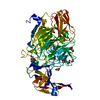
 MEMBRANE PROTEIN / NorC /
  Function and homology information

 Authors
 India, 2items
  Citation
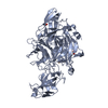
 Structure visualization
 Downloads & links
 Other downloads


 PDBj
PDBj


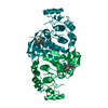
 Assembly


 Mass: 65.409 Da / Num. of mol.: 2 / Source method: obtained synthetically / Formula: Zn
 Sample preparation
 / Beamline: 24-ID-C / Wavelength: 0.97918 Å
 Processing