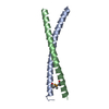
 Movie
Movie Controller


+ Open data

- Basic information
| Entry | Database: PDB / ID: 7d2e | |||||||||
|---|---|---|---|---|---|---|---|---|---|---|
| Title | Tetrameric coiled-coil structure of liprin-alpha2_H3 | |||||||||
 Components Components | Liprin-alpha-2 | |||||||||
 Keywords Keywords |  PROTEIN BINDING / Coiled-coil PROTEIN BINDING / Coiled-coil | |||||||||
| Function / homology |  Function and homology information Function and homology informationstructural constituent of presynapse / dense core granule cytoskeletal transport / Receptor-type tyrosine-protein phosphatases / Acetylcholine Neurotransmitter Release Cycle / postsynaptic specialization / Serotonin Neurotransmitter Release Cycle / regulation of dendritic spine development / Dopamine Neurotransmitter Release Cycle / Norepinephrine Neurotransmitter Release Cycle / Glutamate Neurotransmitter Release Cycle ...structural constituent of presynapse / dense core granule cytoskeletal transport / Receptor-type tyrosine-protein phosphatases / Acetylcholine Neurotransmitter Release Cycle / postsynaptic specialization / Serotonin Neurotransmitter Release Cycle / regulation of dendritic spine development / Dopamine Neurotransmitter Release Cycle / Norepinephrine Neurotransmitter Release Cycle / Glutamate Neurotransmitter Release Cycle / regulation of dendritic spine morphogenesis / regulation of synaptic vesicle exocytosis / presynaptic active zone / cell-matrix adhesion / synapse organization / synaptic vesicle / presynaptic membrane / dendritic spine / axon / glutamatergic synapse / cell surface / extracellular exosome / cytosol / cytoplasmSimilarity search - Function | |||||||||
| Biological species |  Homo sapiens (human) Homo sapiens (human) | |||||||||
| Method | X-RAY DIFFRACTION / SYNCHROTRON / SAD / Resolution: 1.7 Å | |||||||||
 Authors Authors | Liang, M. / Wei, Z. | |||||||||
| Funding support |  China, 2items China, 2items
| |||||||||
 Citation Citation | Journal: Cell Rep / Year: 2021 Title: Oligomerized liprin-alpha promotes phase separation of ELKS for compartmentalization of presynaptic active zone proteins. Authors: Liang, M. / Jin, G. / Xie, X. / Zhang, W. / Li, K. / Niu, F. / Yu, C. / Wei, Z. | |||||||||
| History |
|
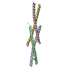
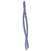
- Structure visualization
| Structure viewer | Molecule: MolmilJmol/JSmol |
|---|
- Downloads & links
-Download
| PDBx/mmCIF format | 7d2e.cif.gz | 114.4 KB | Display | PDBx/mmCIF format |
|---|---|---|---|---|
| PDB format | pdb7d2e.ent.gz | 89.8 KB | Display | PDB format |
| PDBx/mmJSON format | 7d2e.json.gz | Tree view | PDBx/mmJSON format | |
| Others |  Other downloads Other downloads |
-Validation report
| Arichive directory | https://data.pdbj.org/pub/pdb/validation_reports/d2/7d2eftp://data.pdbj.org/pub/pdb/validation_reports/d2/7d2e | HTTPS FTP |
|---|
-Related structure data

-Links
 PDBj
PDBj
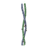
- Assembly
| Deposited unit | 
| ||||||||
|---|---|---|---|---|---|---|---|---|---|
| 1 |
| ||||||||
| Unit cell |
|
-Components
| #1: Protein | Mass: 8665.760 Da / Num. of mol.: 4 / Fragment: UNP residues 164-235 Source method: isolated from a genetically manipulated source Source: (gene. exp.) Homo sapiens (human) / Gene: PPFIA2 / Plasmid: pET32m / Production host:  Escherichia coli BL21(DE3) (bacteria) / Strain (production host): BL21(DE3) / References: UniProt: O75334 Escherichia coli BL21(DE3) (bacteria) / Strain (production host): BL21(DE3) / References: UniProt: O75334#2: Water | ChemComp-HOH / | Water Mass: 18.015 Da / Num. of mol.: 123 / Source method: isolated from a natural source / Formula: H2O Mass: 18.015 Da / Num. of mol.: 123 / Source method: isolated from a natural source / Formula: H2O |
|---|
-Experimental details
-Experiment
| Experiment | Method: X-RAY DIFFRACTION / Number of used crystals: 1 |
|---|
- Sample preparation
| Crystal | Density Matthews: 2.05 Å3/Da / Density % sol: 39.9 % |
|---|---|
| Crystal grow | Temperature: 289 K / Method: vapor diffusion, sitting drop Details: 0.1 M Sodium citrate tribasic dihydrate pH 5.0, 10% w/v Polyethylene glycol 6,000 |
-Data collection
| Diffraction | Mean temperature: 100 K / Serial crystal experiment: N | |||||||||||||||||||||||||||||||||||||||||||||||||||||||||||||||||||||||||||||||||||||||||||||||||||||||||||||||||||||||||||||||||||||||||||||||||||||||||||||||||||||||||||||||||||||||||||||
|---|---|---|---|---|---|---|---|---|---|---|---|---|---|---|---|---|---|---|---|---|---|---|---|---|---|---|---|---|---|---|---|---|---|---|---|---|---|---|---|---|---|---|---|---|---|---|---|---|---|---|---|---|---|---|---|---|---|---|---|---|---|---|---|---|---|---|---|---|---|---|---|---|---|---|---|---|---|---|---|---|---|---|---|---|---|---|---|---|---|---|---|---|---|---|---|---|---|---|---|---|---|---|---|---|---|---|---|---|---|---|---|---|---|---|---|---|---|---|---|---|---|---|---|---|---|---|---|---|---|---|---|---|---|---|---|---|---|---|---|---|---|---|---|---|---|---|---|---|---|---|---|---|---|---|---|---|---|---|---|---|---|---|---|---|---|---|---|---|---|---|---|---|---|---|---|---|---|---|---|---|---|---|---|---|---|---|---|---|---|---|
| Diffraction source | Source: SYNCHROTRON / Site: SSRF / Beamline: BL17U1 / Wavelength: 0.98 Å | |||||||||||||||||||||||||||||||||||||||||||||||||||||||||||||||||||||||||||||||||||||||||||||||||||||||||||||||||||||||||||||||||||||||||||||||||||||||||||||||||||||||||||||||||||||||||||||
| Detector | Type: ADSC QUANTUM 315r / Detector: CCD / Date: Jun 20, 2015 | |||||||||||||||||||||||||||||||||||||||||||||||||||||||||||||||||||||||||||||||||||||||||||||||||||||||||||||||||||||||||||||||||||||||||||||||||||||||||||||||||||||||||||||||||||||||||||||
| Radiation | Protocol: SINGLE WAVELENGTH / Monochromatic (M) / Laue (L): M / Scattering type: x-ray | |||||||||||||||||||||||||||||||||||||||||||||||||||||||||||||||||||||||||||||||||||||||||||||||||||||||||||||||||||||||||||||||||||||||||||||||||||||||||||||||||||||||||||||||||||||||||||||
| Radiation wavelength | Wavelength: 0.98 Å / Relative weight: 1 | |||||||||||||||||||||||||||||||||||||||||||||||||||||||||||||||||||||||||||||||||||||||||||||||||||||||||||||||||||||||||||||||||||||||||||||||||||||||||||||||||||||||||||||||||||||||||||||
| Reflection | Resolution: 1.7→50 Å / Num. obs: 30192 / % possible obs: 98.4 % / Redundancy: 3.7 % / Biso Wilson estimate: 29.34 Å2 / Rmerge(I) obs: 0.037 / Rpim(I) all: 0.022 / Rrim(I) all: 0.043 / Χ2: 0.903 / Net I/σ(I): 15.6 | |||||||||||||||||||||||||||||||||||||||||||||||||||||||||||||||||||||||||||||||||||||||||||||||||||||||||||||||||||||||||||||||||||||||||||||||||||||||||||||||||||||||||||||||||||||||||||||
| Reflection shell | Diffraction-ID: 1
|
- Processing
| Software |
| |||||||||||||||||||||||||||||||||||||||||||||||||||||||||||||||||||||||||||||||||||||||||||||||||||||||||||||||||||||||||||||
|---|---|---|---|---|---|---|---|---|---|---|---|---|---|---|---|---|---|---|---|---|---|---|---|---|---|---|---|---|---|---|---|---|---|---|---|---|---|---|---|---|---|---|---|---|---|---|---|---|---|---|---|---|---|---|---|---|---|---|---|---|---|---|---|---|---|---|---|---|---|---|---|---|---|---|---|---|---|---|---|---|---|---|---|---|---|---|---|---|---|---|---|---|---|---|---|---|---|---|---|---|---|---|---|---|---|---|---|---|---|---|---|---|---|---|---|---|---|---|---|---|---|---|---|---|---|---|
| Refinement | Method to determine structure: SAD / Resolution: 1.7→50 Å / SU ML: 0.22 / Cross valid method: THROUGHOUT / σ(F): 1.35 / Phase error: 28.84 / Stereochemistry target values: ML
| |||||||||||||||||||||||||||||||||||||||||||||||||||||||||||||||||||||||||||||||||||||||||||||||||||||||||||||||||||||||||||||
| Solvent computation | Shrinkage radii: 0.9 Å / VDW probe radii: 1.11 Å / Solvent model: FLAT BULK SOLVENT MODEL | |||||||||||||||||||||||||||||||||||||||||||||||||||||||||||||||||||||||||||||||||||||||||||||||||||||||||||||||||||||||||||||
| Displacement parameters | Biso max: 118.47 Å2 / Biso mean: 46.1563 Å2 / Biso min: 16.58 Å2 | |||||||||||||||||||||||||||||||||||||||||||||||||||||||||||||||||||||||||||||||||||||||||||||||||||||||||||||||||||||||||||||
| Refinement step | Cycle: final / Resolution: 1.7→50 Å
| |||||||||||||||||||||||||||||||||||||||||||||||||||||||||||||||||||||||||||||||||||||||||||||||||||||||||||||||||||||||||||||
| Refine LS restraints |
| |||||||||||||||||||||||||||||||||||||||||||||||||||||||||||||||||||||||||||||||||||||||||||||||||||||||||||||||||||||||||||||
| LS refinement shell | Refine-ID: X-RAY DIFFRACTION / Rfactor Rfree error: 0
| |||||||||||||||||||||||||||||||||||||||||||||||||||||||||||||||||||||||||||||||||||||||||||||||||||||||||||||||||||||||||||||
| Refinement TLS params. | Method: refined / Refine-ID: X-RAY DIFFRACTION
| |||||||||||||||||||||||||||||||||||||||||||||||||||||||||||||||||||||||||||||||||||||||||||||||||||||||||||||||||||||||||||||
| Refinement TLS group |
|
