 Movie
Movie Controller


+ Open data

- Basic information
| Entry | Database: PDB / ID: 7d0m | |||||||||
|---|---|---|---|---|---|---|---|---|---|---|
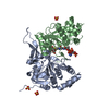
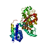
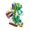
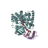
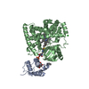
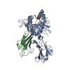
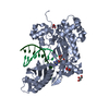
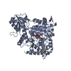
| Title | Crystal structure of mouse CRY1 with bound cryoprotectant | |||||||||
 Components Components | Cryptochrome-1 | |||||||||
 Keywords Keywords | CIRCADIAN CLOCK PROTEIN / CRY / CRY1 / cryptochrome / PHR | |||||||||
| Function / homology |  Function and homology information Function and homology informationnegative regulation of glucocorticoid secretion / negative regulation of glucocorticoid receptor signaling pathway / negative regulation of circadian rhythm / negative regulation of G protein-coupled receptor signaling pathway / lipid storage / regulation of DNA damage checkpoint / response to glucagon / regulation of gluconeogenesis / entrainment of circadian clock by photoperiod / E-box binding ...negative regulation of glucocorticoid secretion / negative regulation of glucocorticoid receptor signaling pathway / negative regulation of circadian rhythm / negative regulation of G protein-coupled receptor signaling pathway / lipid storage / regulation of DNA damage checkpoint / response to glucagon / regulation of gluconeogenesis / entrainment of circadian clock by photoperiod / E-box binding / photoreceptor activity / response to light stimulus / phosphatase binding / negative regulation of gluconeogenesis / signal transduction in response to DNA damage / positive regulation of gluconeogenesis / negative regulation of protein ubiquitination / FAD binding / response to activity / positive regulation of protein ubiquitination / gluconeogenesis / nuclear receptor binding / circadian regulation of gene expression / response to insulin / regulation of circadian rhythm / kinase binding / circadian rhythm / histone deacetylase binding / glucose homeostasis / double-stranded DNA binding / DNA-binding transcription factor binding / negative regulation of DNA-templated transcription / protein kinase binding / negative regulation of transcription by RNA polymerase II / mitochondrion / DNA binding / nucleoplasm / nucleus / cytosol / cytoplasmSimilarity search - Function | |||||||||
| Biological species |  Mus musculus (house mouse) Mus musculus (house mouse) | |||||||||
| Method | X-RAY DIFFRACTION / SYNCHROTRON / MOLECULAR REPLACEMENT / Resolution: 1.95 Å | |||||||||
 Authors Authors | Miller, S.A. / Aikawa, Y. / Hirota, T. | |||||||||
| Funding support |  Japan, 2items Japan, 2items
| |||||||||
 Citation Citation | Journal: Proc.Natl.Acad.Sci.USA / Year: 2021 Title: Structural differences in the FAD-binding pockets and lid loops of mammalian CRY1 and CRY2 for isoform-selective regulation. Authors: Miller, S. / Srivastava, A. / Nagai, Y. / Aikawa, Y. / Tama, F. / Hirota, T. | |||||||||
| History |
|
- Structure visualization
| Structure viewer | Molecule: MolmilJmol/JSmol |
|---|
- Downloads & links
-Download
| PDBx/mmCIF format | 7d0m.cif.gz | 254.9 KB | Display | PDBx/mmCIF format |
|---|---|---|---|---|
| PDB format | pdb7d0m.ent.gz | 166 KB | Display | PDB format |
| PDBx/mmJSON format | 7d0m.json.gz | Tree view | PDBx/mmJSON format | |
| Others |  Other downloads Other downloads |
-Validation report
| Arichive directory | https://data.pdbj.org/pub/pdb/validation_reports/d0/7d0mftp://data.pdbj.org/pub/pdb/validation_reports/d0/7d0m | HTTPS FTP |
|---|
-Related structure data
| Related structure data |  7d0nC 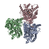 7dliC 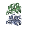 7ej9C 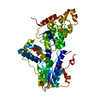 6kx4S S: Starting model for refinement C: citing same article ( |
|---|---|
| Similar structure data |


-Links
 PDBj
PDBj

- Assembly
| Deposited unit | 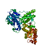
| ||||||||||||
|---|---|---|---|---|---|---|---|---|---|---|---|---|---|
| 1 |
| ||||||||||||
| Unit cell |
|
-Components
| #1: Protein | Mass: 57371.734 Da / Num. of mol.: 1 Source method: isolated from a genetically manipulated source Source: (gene. exp.) Mus musculus (house mouse) / Gene: Cry1 / Production host:   Spodoptera frugiperda (fall armyworm) / References: UniProt: P97784 Spodoptera frugiperda (fall armyworm) / References: UniProt: P97784 |
|---|---|
| #2: Chemical | ChemComp-PG4 / Polyethylene glycol  Mass: 194.226 Da / Num. of mol.: 1 / Source method: obtained synthetically / Formula: C8H18O5 / Comment: precipitant*YM Mass: 194.226 Da / Num. of mol.: 1 / Source method: obtained synthetically / Formula: C8H18O5 / Comment: precipitant*YM |
| #3: Chemical | ChemComp-PEG / Diethylene glycol  Mass: 106.120 Da / Num. of mol.: 1 / Source method: obtained synthetically / Formula: C4H10O3 Mass: 106.120 Da / Num. of mol.: 1 / Source method: obtained synthetically / Formula: C4H10O3 |
| #4: Water | ChemComp-HOH / Water Mass: 18.015 Da / Num. of mol.: 445 / Source method: isolated from a natural source / Formula: H2O Mass: 18.015 Da / Num. of mol.: 445 / Source method: isolated from a natural source / Formula: H2O |
| Has ligand of interest | N |
-Experimental details
-Experiment
| Experiment | Method: X-RAY DIFFRACTION / Number of used crystals: 1 |
|---|
- Sample preparation
| Crystal | Density Matthews: 2.05 Å3/Da / Density % sol: 40.02 % |
|---|---|
| Crystal grow | Temperature: 293 K / Method: vapor diffusion, hanging drop Details: 0.25 M NH4Cl, 22% w/v PEG 3350, 3% v/v ethylene glycol |
-Data collection
| Diffraction | Mean temperature: 100 K / Serial crystal experiment: N |
|---|---|
| Diffraction source | Source: SYNCHROTRON / Site: Photon Factory / Beamline: BL-17A / Wavelength: 0.98 Å |
| Detector | Type: DECTRIS EIGER X 16M / Detector: PIXEL / Date: Feb 23, 2017 |
| Radiation | Protocol: SINGLE WAVELENGTH / Monochromatic (M) / Laue (L): M / Scattering type: x-ray |
| Radiation wavelength | Wavelength: 0.98 Å / Relative weight: 1 |
| Reflection | Resolution: 1.95→67.25 Å / Num. obs: 35031 / % possible obs: 99.5 % / Redundancy: 6 % / Biso Wilson estimate: 14.94 Å2 / CC1/2: 0.994 / Rmerge(I) obs: 0.139 / Rpim(I) all: 0.062 / Rrim(I) all: 0.153 / Net I/σ(I): 8.2 |
| Reflection shell | Resolution: 1.95→2.02 Å / Redundancy: 6.1 % / Rmerge(I) obs: 0.496 / Mean I/σ(I) obs: 3.2 / Num. unique obs: 5000 / CC1/2: 0.908 / Rpim(I) all: 0.22 / Rrim(I) all: 0.544 / % possible all: 99.2 |
- Processing
| Software |
| ||||||||||||||||||||||||||||||||||||||||||||||||||||||||||||||||||||||||||||||||||||||||||||||||||
|---|---|---|---|---|---|---|---|---|---|---|---|---|---|---|---|---|---|---|---|---|---|---|---|---|---|---|---|---|---|---|---|---|---|---|---|---|---|---|---|---|---|---|---|---|---|---|---|---|---|---|---|---|---|---|---|---|---|---|---|---|---|---|---|---|---|---|---|---|---|---|---|---|---|---|---|---|---|---|---|---|---|---|---|---|---|---|---|---|---|---|---|---|---|---|---|---|---|---|---|
| Refinement | Method to determine structure: MOLECULAR REPLACEMENT Starting model: 6KX4 Resolution: 1.95→67.25 Å / SU ML: 0.1769 / Cross valid method: FREE R-VALUE / σ(F): 1.34 / Phase error: 19.7862 Stereochemistry target values: GeoStd + Monomer Library + CDL v1.2
| ||||||||||||||||||||||||||||||||||||||||||||||||||||||||||||||||||||||||||||||||||||||||||||||||||
| Solvent computation | Shrinkage radii: 0.9 Å / VDW probe radii: 1.11 Å / Solvent model: FLAT BULK SOLVENT MODEL | ||||||||||||||||||||||||||||||||||||||||||||||||||||||||||||||||||||||||||||||||||||||||||||||||||
| Displacement parameters | Biso mean: 15.55 Å2 | ||||||||||||||||||||||||||||||||||||||||||||||||||||||||||||||||||||||||||||||||||||||||||||||||||
| Refinement step | Cycle: LAST / Resolution: 1.95→67.25 Å
| ||||||||||||||||||||||||||||||||||||||||||||||||||||||||||||||||||||||||||||||||||||||||||||||||||
| Refine LS restraints |
| ||||||||||||||||||||||||||||||||||||||||||||||||||||||||||||||||||||||||||||||||||||||||||||||||||
| LS refinement shell |
| ||||||||||||||||||||||||||||||||||||||||||||||||||||||||||||||||||||||||||||||||||||||||||||||||||
| Refinement TLS params. | Method: refined / Refine-ID: X-RAY DIFFRACTION
| ||||||||||||||||||||||||||||||||||||||||||||||||||||||||||||||||||||||||||||||||||||||||||||||||||
| Refinement TLS group | Refine-ID: X-RAY DIFFRACTION / Auth asym-ID: A / Label asym-ID: A
|
