 Movie
Movie Controller

[English] 日本語
 Yorodumi
- PDB-7cu5: N-Glycosylation of PD-1 and glycosylation dependent binding of PD... -
+ Open data

- Basic information
| Entry | Database: PDB / ID: 7cu5 | ||||||
|---|---|---|---|---|---|---|---|
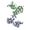
| Title | N-Glycosylation of PD-1 and glycosylation dependent binding of PD-1 specific monoclonal antibody camrelizumab | ||||||
 Components Components |
| ||||||
 Keywords Keywords |  IMMUNE SYSTEM / Camrelizumab / glycosylation / PD-1. IMMUNE SYSTEM / Camrelizumab / glycosylation / PD-1. | ||||||
| Function / homology |  Function and homology information Function and homology informationnegative regulation of tolerance induction / regulatory T cell apoptotic process / negative regulation of immune response / B cell apoptotic process / positive regulation of T cell apoptotic process / negative regulation of B cell apoptotic process / humoral immune response / PD-1 signaling / regulation of immune response / Potential therapeutics for SARS ...negative regulation of tolerance induction / regulatory T cell apoptotic process / negative regulation of immune response / B cell apoptotic process / positive regulation of T cell apoptotic process / negative regulation of B cell apoptotic process / humoral immune response / PD-1 signaling / regulation of immune response / Potential therapeutics for SARS / adaptive immune response / external side of plasma membrane / apoptotic process / plasma membraneSimilarity search - Function | ||||||
| Biological species |  Homo sapiens (human) Homo sapiens (human) | ||||||
| Method | X-RAY DIFFRACTION / SYNCHROTRON / MOLECULAR REPLACEMENT / Resolution: 2.81 Å | ||||||
 Authors Authors | Liu, K.F. / Tan, S.G. / Jin, W.J. / Guan, J.W. / Wang, W.L. / Sun, H. / Qi, J.X. / Yan, J.H. / Chai, Y. / Wang, Z.F. ...Liu, K.F. / Tan, S.G. / Jin, W.J. / Guan, J.W. / Wang, W.L. / Sun, H. / Qi, J.X. / Yan, J.H. / Chai, Y. / Wang, Z.F. / Chu, X.D. / Gao, G.F. | ||||||
 Citation Citation | Journal: Embo Rep. / Year: 2020 Title: N-glycosylation of PD-1 promotes binding of camrelizumab. Authors: Liu, K. / Tan, S. / Jin, W. / Guan, J. / Wang, Q. / Sun, H. / Qi, J. / Yan, J. / Chai, Y. / Wang, Z. / Deng, C. / Gao, G.F. | ||||||
| History |
|
- Structure visualization
| Structure viewer | Molecule: MolmilJmol/JSmol |
|---|
- Downloads & links
-Download
| PDBx/mmCIF format | 7cu5.cif.gz | 162.5 KB | Display | PDBx/mmCIF format |
|---|---|---|---|---|
| PDB format | pdb7cu5.ent.gz | 116.4 KB | Display | PDB format |
| PDBx/mmJSON format | 7cu5.json.gz | Tree view | PDBx/mmJSON format | |
| Others |  Other downloads Other downloads |
-Validation report
| Arichive directory | https://data.pdbj.org/pub/pdb/validation_reports/cu/7cu5ftp://data.pdbj.org/pub/pdb/validation_reports/cu/7cu5 | HTTPS FTP |
|---|
-Related structure data
| Related structure data |  5wt9S S: Starting model for refinement |
|---|---|
| Similar structure data |

-Links
 PDBj
PDBj


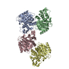
- Assembly
| Deposited unit | 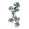
| ||||||||||||
|---|---|---|---|---|---|---|---|---|---|---|---|---|---|
| 1 | 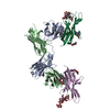
| ||||||||||||
| Unit cell |
|
-Components
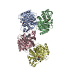
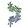
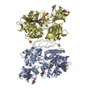
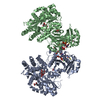
-Antibody / Protein , 2 types, 4 molecules BAEQ
| #1: Antibody | Mass: 24791.680 Da / Num. of mol.: 2 Source method: isolated from a genetically manipulated source Source: (gene. exp.) Homo sapiens (human) / Production host:  Escherichia coli (E. coli) Escherichia coli (E. coli)#2: Protein | / hPD-1Mass: 13327.914 Da / Num. of mol.: 2 Source method: isolated from a genetically manipulated source Source: (gene. exp.) Homo sapiens (human) / Gene: PDCD1, PD1 / Cell line (production host): HEK293 / Production host: Homo sapiens (human) / References: UniProt: Q15116 |
|---|
-Sugars , 4 types, 7 molecules 
| #3: Polysaccharide | alpha-D-mannopyranose-(1-6)-alpha-D-mannopyranose-(1-4)-2-acetamido-2-deoxy-beta-D-glucopyranose-(1- ...alpha-D-mannopyranose-(1-6)-alpha-D-mannopyranose-(1-4)-2-acetamido-2-deoxy-beta-D-glucopyranose-(1-4)-[alpha-L-fucopyranose-(1-6)]2-acetamido-2-deoxy-beta-D-glucopyranose / Mass: 894.823 Da / Num. of mol.: 1 Source method: isolated from a genetically manipulated source | ||||
|---|---|---|---|---|---|
| #4: Polysaccharide | / Mass: 570.542 Da / Num. of mol.: 2 Source method: isolated from a genetically manipulated source #5: Polysaccharide | alpha-D-mannopyranose-(1-3)-alpha-D-mannopyranose-(1-4)-2-acetamido-2-deoxy-beta-D-glucopyranose-(1- ...alpha-D-mannopyranose-(1-3)-alpha-D-mannopyranose-(1-4)-2-acetamido-2-deoxy-beta-D-glucopyranose-(1-4)-[alpha-L-fucopyranose-(1-6)]2-acetamido-2-deoxy-beta-D-glucopyranose | / Mass: 894.823 Da / Num. of mol.: 1Source method: isolated from a genetically manipulated source #6: Sugar | N-Acetylglucosamine Type: D-saccharide, beta linking / Mass: 221.208 Da / Num. of mol.: 3 Type: D-saccharide, beta linking / Mass: 221.208 Da / Num. of mol.: 3Source method: isolated from a genetically manipulated source Formula: C8H15NO6 |
-Details
| Has ligand of interest | N |
|---|
-Experimental details
-Experiment
| Experiment | Method: X-RAY DIFFRACTION / Number of used crystals: 1 |
|---|
- Sample preparation
| Crystal | Density Matthews: 3.86 Å3/Da / Density % sol: 68.14 % |
|---|---|
| Crystal grow | Temperature: 291 K / Method: vapor diffusion, sitting drop Details: 0.1M cadmium chloride, 0.1M Na acetate, pH4.6, 30 %(v/v) PEG 400 a month later. |
-Data collection
| Diffraction | Mean temperature: 100 K / Serial crystal experiment: N |
|---|---|
| Diffraction source | Source: SYNCHROTRON / Site: SSRF  / Beamline: BL19U1 / Wavelength: 0.97918 Å / Beamline: BL19U1 / Wavelength: 0.97918 Å |
| Detector | Type: DECTRIS PILATUS 6M / Detector: PIXEL / Date: May 14, 2019 |
| Radiation | Protocol: SINGLE WAVELENGTH / Monochromatic (M) / Laue (L): M / Scattering type: x-ray |
| Radiation wavelength | Wavelength: 0.97918 Å / Relative weight: 1 |
| Reflection | Resolution: 2.8→50 Å / Num. obs: 27286 / % possible obs: 98.4 % / Redundancy: 3.4 % / Biso Wilson estimate: 34.4 Å2 / Rmerge(I) obs: 0.079 / Net I/σ(I): 11.2 |
| Reflection shell | Resolution: 2.8→2.9 Å / Rmerge(I) obs: 0.692 / Num. unique obs: 2726 |
- Processing
| Software |
| ||||||||||||||||||||||||||||||||||||||||||||||||||||||||||||||||||||||
|---|---|---|---|---|---|---|---|---|---|---|---|---|---|---|---|---|---|---|---|---|---|---|---|---|---|---|---|---|---|---|---|---|---|---|---|---|---|---|---|---|---|---|---|---|---|---|---|---|---|---|---|---|---|---|---|---|---|---|---|---|---|---|---|---|---|---|---|---|---|---|---|
| Refinement | Method to determine structure: MOLECULAR REPLACEMENT Starting model: 5WT9 Resolution: 2.81→29.69 Å / SU ML: 0.3635 / Cross valid method: FREE R-VALUE / σ(F): 1.96 / Phase error: 27.4888 Stereochemistry target values: GeoStd + Monomer Library + CDL v1.2
| ||||||||||||||||||||||||||||||||||||||||||||||||||||||||||||||||||||||
| Solvent computation | Shrinkage radii: 0.9 Å / VDW probe radii: 1.11 Å / Solvent model: FLAT BULK SOLVENT MODEL | ||||||||||||||||||||||||||||||||||||||||||||||||||||||||||||||||||||||
| Displacement parameters | Biso mean: 50.7 Å2 | ||||||||||||||||||||||||||||||||||||||||||||||||||||||||||||||||||||||
| Refinement step | Cycle: LAST / Resolution: 2.81→29.69 Å
| ||||||||||||||||||||||||||||||||||||||||||||||||||||||||||||||||||||||
| Refine LS restraints |
| ||||||||||||||||||||||||||||||||||||||||||||||||||||||||||||||||||||||
| LS refinement shell |
|
