 Movie
Movie Controller

[English] 日本語
 Yorodumi
- PDB-7csj: Aminoglycoside 2'-N-acetyltransferase from Mycolicibacterium smeg... -
+ Open data

- Basic information
| Entry | Database: PDB / ID: 7csj | ||||||
|---|---|---|---|---|---|---|---|
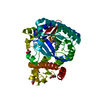
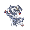
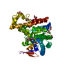
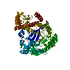
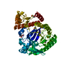
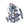
| Title | Aminoglycoside 2'-N-acetyltransferase from Mycolicibacterium smegmatis-Complex with Coenzyme A and Gentamicin | ||||||
 Components Components | Aminoglycoside 2'-N-acetyltransferase | ||||||
 Keywords Keywords |  TRANSFERASE / aminoglycoside acetyltransferase / antibiotics / gentamicin TRANSFERASE / aminoglycoside acetyltransferase / antibiotics / gentamicin | ||||||
| Function / homology |  Function and homology information Function and homology informationacyltransferase activity, transferring groups other than amino-acyl groups / Transferases; Acyltransferases; Transferring groups other than aminoacyl groups / response to antibioticSimilarity search - Function | ||||||
| Biological species |  Mycolicibacterium smegmatis (bacteria) Mycolicibacterium smegmatis (bacteria) | ||||||
| Method | X-RAY DIFFRACTION / SYNCHROTRON / MOLECULAR REPLACEMENT / Resolution: 2.168 Å | ||||||
 Authors Authors | Jeong, C.S. / Hwang, J. / Do, H. / Lee, J.H. | ||||||
 Citation Citation | Journal: Sci Rep / Year: 2020 Title: Structural and biochemical analyses of an aminoglycoside 2'-N-acetyltransferase from Mycolicibacterium smegmatis. Authors: Jeong, C.S. / Hwang, J. / Do, H. / Cha, S.S. / Oh, T.J. / Kim, H.J. / Park, H.H. / Lee, J.H. | ||||||
| History |
|
- Structure visualization
| Structure viewer | Molecule: MolmilJmol/JSmol |
|---|
- Downloads & links
-Download
| PDBx/mmCIF format | 7csj.cif.gz | 92.4 KB | Display | PDBx/mmCIF format |
|---|---|---|---|---|
| PDB format | pdb7csj.ent.gz | 68.5 KB | Display | PDB format |
| PDBx/mmJSON format | 7csj.json.gz | Tree view | PDBx/mmJSON format | |
| Others |  Other downloads Other downloads |
-Validation report
| Arichive directory | https://data.pdbj.org/pub/pdb/validation_reports/cs/7csjftp://data.pdbj.org/pub/pdb/validation_reports/cs/7csj | HTTPS FTP |
|---|
-Related structure data
| Related structure data |  7crmSC 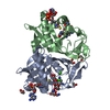 7cs0C 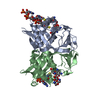 7cs1C  7csiC S: Starting model for refinement C: citing same article ( |
|---|---|
| Similar structure data |
-Links
 PDBj
PDBj


- Assembly
| Deposited unit | 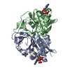
| ||||||||
|---|---|---|---|---|---|---|---|---|---|
| 1 |
| ||||||||
| Unit cell |
|
-Components
| #1: Protein | Mass: 23198.838 Da / Num. of mol.: 2 Source method: isolated from a genetically manipulated source Source: (gene. exp.) Mycolicibacterium smegmatis (strain ATCC 700084 / mc(2)155) (bacteria)Gene: aac, MSMEG_0434, MSMEI_0423 / Production host: Escherichia coli (E. coli)References: UniProt: P94968, Transferases; Acyltransferases; Transferring groups other than aminoacyl groups#2: Chemical | ChemComp-51G / |   Mass: 477.595 Da / Num. of mol.: 1 / Source method: obtained synthetically / Formula: C21H43N5O7 Mass: 477.595 Da / Num. of mol.: 1 / Source method: obtained synthetically / Formula: C21H43N5O7#3: Chemical | Coenzyme A  Mass: 767.534 Da / Num. of mol.: 2 / Source method: obtained synthetically / Formula: C21H36N7O16P3S Mass: 767.534 Da / Num. of mol.: 2 / Source method: obtained synthetically / Formula: C21H36N7O16P3S#4: Water | ChemComp-HOH / | Water Mass: 18.015 Da / Num. of mol.: 113 / Source method: isolated from a natural source / Formula: H2O Mass: 18.015 Da / Num. of mol.: 113 / Source method: isolated from a natural source / Formula: H2OHas ligand of interest | N | |
|---|
-Experimental details
-Experiment
| Experiment | Method: X-RAY DIFFRACTION / Number of used crystals: 1 |
|---|
- Sample preparation
| Crystal | Density Matthews: 2.07 Å3/Da / Density % sol: 40.51 % |
|---|---|
| Crystal grow | Temperature: 295 K / Method: vapor diffusion, sitting drop Details: 0.1M sodium citrate: citric acid (pH 5.5), 20% (w/v) PEG 3000 |
-Data collection
| Diffraction | Mean temperature: 100 K / Serial crystal experiment: N |
|---|---|
| Diffraction source | Source: SYNCHROTRON / Site: PAL/PLS  / Beamline: 5C (4A) / Wavelength: 1 Å / Beamline: 5C (4A) / Wavelength: 1 Å |
| Detector | Type: DECTRIS EIGER X 9M / Detector: PIXEL / Date: Oct 22, 2019 |
| Radiation | Protocol: SINGLE WAVELENGTH / Monochromatic (M) / Laue (L): M / Scattering type: x-ray |
| Radiation wavelength | Wavelength: 1 Å / Relative weight: 1 |
| Reflection | Resolution: 2.168→50 Å / Num. obs: 20679 / % possible obs: 97.1 % / Redundancy: 5.6 % / Rmerge(I) obs: 0.078 / Net I/σ(I): 37.985 |
| Reflection shell | Resolution: 2.17→2.21 Å / Redundancy: 4.9 % / Rmerge(I) obs: 0.325 / Mean I/σ(I) obs: 4.46 / Num. unique obs: 791 / % possible all: 76.5 |
- Processing
| Software |
| |||||||||||||||||||||||||||||||||||||||||||||||||
|---|---|---|---|---|---|---|---|---|---|---|---|---|---|---|---|---|---|---|---|---|---|---|---|---|---|---|---|---|---|---|---|---|---|---|---|---|---|---|---|---|---|---|---|---|---|---|---|---|---|---|
| Refinement | Method to determine structure: MOLECULAR REPLACEMENT Starting model: 7CRM Resolution: 2.168→41.95 Å / SU ML: 0.27 / Cross valid method: FREE R-VALUE / σ(F): 1.35 / Phase error: 25.28 / Stereochemistry target values: ML
| |||||||||||||||||||||||||||||||||||||||||||||||||
| Solvent computation | Shrinkage radii: 0.9 Å / VDW probe radii: 1.11 Å / Solvent model: FLAT BULK SOLVENT MODEL | |||||||||||||||||||||||||||||||||||||||||||||||||
| Refinement step | Cycle: LAST / Resolution: 2.168→41.95 Å
| |||||||||||||||||||||||||||||||||||||||||||||||||
| Refine LS restraints |
| |||||||||||||||||||||||||||||||||||||||||||||||||
| LS refinement shell |
|
