 Movie
Movie Controller


+ Open data

- Basic information
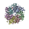
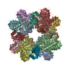
| Entry | Database: PDB / ID: 7cpr | ||||||
|---|---|---|---|---|---|---|---|
| Title | glutamine synthetase from Drosophila | ||||||
 Components Components | Glutamine synthetase 2 cytoplasmic | ||||||
 Keywords Keywords |  LIGASE / complex LIGASE / complex | ||||||
| Function / homology |  Function and homology information Function and homology informationAstrocytic Glutamate-Glutamine Uptake And Metabolism / : / Glutamate and glutamine metabolism / glutamate catabolic process / glutamine synthetase / glutamine biosynthetic process / glutamine synthetase activity / synapse assembly / ATP binding / cytoplasmSimilarity search - Function | ||||||
| Biological species |  Drosophila melanogaster (fruit fly) Drosophila melanogaster (fruit fly) | ||||||
| Method | X-RAY DIFFRACTION / SYNCHROTRON / MOLECULAR REPLACEMENT / Resolution: 2.12 Å | ||||||
 Authors Authors | Yin, H.S. / Chen, W.T. | ||||||
| Funding support |  Taiwan, 1items Taiwan, 1items
| ||||||
 Citation Citation | Journal: Biomolecules / Year: 2020 Title: Structural Insight into the Contributions of the N-Terminus and Key Active-Site Residues to the Catalytic Efficiency of Glutamine Synthetase 2. Authors: Chen, W.T. / Yang, H.Y. / Lin, C.Y. / Lee, Y.Z. / Ma, S.C. / Chen, W.C. / Yin, H.S. | ||||||
| History |
|
- Structure visualization
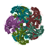
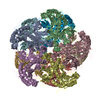
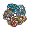
| Structure viewer | Molecule: MolmilJmol/JSmol |
|---|
- Downloads & links
-Download
| PDBx/mmCIF format | 7cpr.cif.gz | 715.2 KB | Display | PDBx/mmCIF format |
|---|---|---|---|---|
| PDB format | pdb7cpr.ent.gz | 593.6 KB | Display | PDB format |
| PDBx/mmJSON format | 7cpr.json.gz | Tree view | PDBx/mmJSON format | |
| Others |  Other downloads Other downloads |
-Validation report
| Arichive directory | https://data.pdbj.org/pub/pdb/validation_reports/cp/7cprftp://data.pdbj.org/pub/pdb/validation_reports/cp/7cpr | HTTPS FTP |
|---|
-Related structure data
| Related structure data | 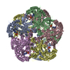 2ojwS S: Starting model for refinement |
|---|---|
| Similar structure data |

-Links
 PDBj
PDBj
- Assembly
| Deposited unit | 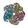
| ||||||||
|---|---|---|---|---|---|---|---|---|---|
| 1 |
| ||||||||
| Unit cell |
|
-Components
| #1: Protein | Mass: 41136.020 Da / Num. of mol.: 10 Source method: isolated from a genetically manipulated source Source: (gene. exp.) Drosophila melanogaster (fruit fly) / Gene: Gs2, CG1743 / Production host:  Escherichia coli (E. coli) / References: UniProt: P20478, glutamine synthetase Escherichia coli (E. coli) / References: UniProt: P20478, glutamine synthetase#2: Chemical | ChemComp-ADP / Adenosine diphosphate  Mass: 427.201 Da / Num. of mol.: 10 / Source method: obtained synthetically / Formula: C10H15N5O10P2 / Feature type: SUBJECT OF INVESTIGATION / Comment: ADP, energy-carrying molecule*YM Mass: 427.201 Da / Num. of mol.: 10 / Source method: obtained synthetically / Formula: C10H15N5O10P2 / Feature type: SUBJECT OF INVESTIGATION / Comment: ADP, energy-carrying molecule*YM#3: Water | ChemComp-HOH / | Water Mass: 18.015 Da / Num. of mol.: 520 / Source method: isolated from a natural source / Formula: H2O Mass: 18.015 Da / Num. of mol.: 520 / Source method: isolated from a natural source / Formula: H2OHas ligand of interest | Y | |
|---|
-Experimental details
-Experiment
| Experiment | Method: X-RAY DIFFRACTION / Number of used crystals: 1 |
|---|
- Sample preparation
| Crystal | Density Matthews: 2.57 Å3/Da / Density % sol: 52.09 % |
|---|---|
| Crystal grow | Temperature: 293 K / Method: vapor diffusion, hanging drop Details: 20 mM Tris-HCl, pH 7.9, 150 mM NaCl, 5mM MgCl2, 5 mM ATP, 1 mM sodium 2-mercaptoethanesulfonate |
-Data collection
| Diffraction | Mean temperature: 100 K / Serial crystal experiment: N |
|---|---|
| Diffraction source | Source: SYNCHROTRON / Site: NSRRC / Beamline: BL13B1 / Wavelength: 1 Å |
| Detector | Type: MAR CCD 165 mm / Detector: CCD / Date: Oct 23, 2016 |
| Radiation | Protocol: SINGLE WAVELENGTH / Monochromatic (M) / Laue (L): M / Scattering type: x-ray |
| Radiation wavelength | Wavelength: 1 Å / Relative weight: 1 |
| Reflection | Resolution: 2.12→178.68 Å / Num. obs: 234758 / % possible obs: 98 % / Redundancy: 2.8 % / CC1/2: 1 / Net I/σ(I): 16.14 |
| Reflection shell | Resolution: 2.12→2.2 Å / Num. unique obs: 234758 / CC1/2: 1 |
- Processing
| Software |
| ||||||||||||||||||||||||||||||||||||||||||||||||||||||||||||
|---|---|---|---|---|---|---|---|---|---|---|---|---|---|---|---|---|---|---|---|---|---|---|---|---|---|---|---|---|---|---|---|---|---|---|---|---|---|---|---|---|---|---|---|---|---|---|---|---|---|---|---|---|---|---|---|---|---|---|---|---|---|
| Refinement | Method to determine structure: MOLECULAR REPLACEMENT Starting model: 2OJW Resolution: 2.12→178.68 Å / Cor.coef. Fo:Fc: 0.955 / Cor.coef. Fo:Fc free: 0.928 / SU B: 7.125 / SU ML: 0.178 / Cross valid method: THROUGHOUT / σ(F): 0 / ESU R: 0.235 / ESU R Free: 0.197 / Stereochemistry target values: MAXIMUM LIKELIHOOD Details: HYDROGENS HAVE BEEN ADDED IN THE RIDING POSITIONS U VALUES : REFINED INDIVIDUALLY
| ||||||||||||||||||||||||||||||||||||||||||||||||||||||||||||
| Solvent computation | Ion probe radii: 0.8 Å / Shrinkage radii: 0.8 Å / VDW probe radii: 1.2 Å / Solvent model: MASK | ||||||||||||||||||||||||||||||||||||||||||||||||||||||||||||
| Displacement parameters | Biso max: 217.58 Å2 / Biso mean: 51.442 Å2 / Biso min: 18.85 Å2
| ||||||||||||||||||||||||||||||||||||||||||||||||||||||||||||
| Refinement step | Cycle: final / Resolution: 2.12→178.68 Å
| ||||||||||||||||||||||||||||||||||||||||||||||||||||||||||||
| Refine LS restraints |
| ||||||||||||||||||||||||||||||||||||||||||||||||||||||||||||
| LS refinement shell | Resolution: 2.12→2.175 Å / Rfactor Rfree error: 0 / Total num. of bins used: 20
|
