| Entry | Database: PDB / ID: 7cnx
|
|---|
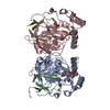
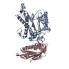
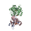
| Title | Crystal structure of Apo PSD from E. coli (2.63 A) |
|---|
 Components Components | - Phosphatidylserine decarboxylase alpha chain
- Phosphatidylserine decarboxylase beta chain
|
|---|
 Keywords Keywords |  LYASE / Phosphatidylserine decarboxylase / pyruvoyl-dependent decarboxylase / auto-cleaved / serine protease / Apo state / MEMBRANE PROTEIN LYASE / Phosphatidylserine decarboxylase / pyruvoyl-dependent decarboxylase / auto-cleaved / serine protease / Apo state / MEMBRANE PROTEIN |
|---|
| Function / homology | phosphatidylserine decarboxylase / Phosphatidylserine decarboxylase-related / Phosphatidylserine decarboxylase / Phosphatidylserine decarboxylase, prokaryotic type 1 / Phosphatidylserine decarboxylase / phosphatidylserine decarboxylase activity / phosphatidylethanolamine biosynthetic process / plasma membrane / Phosphatidylserine decarboxylase proenzyme Function and homology information Function and homology information |
|---|
| Biological species |   Escherichia coli K-12 (bacteria) Escherichia coli K-12 (bacteria) |
|---|
| Method | X-RAY DIFFRACTION / SYNCHROTRON / SAD / Resolution: 2.63 Å |
|---|
 Authors Authors | Kim, J. / Cho, G. |
|---|
| Funding support |  Korea, Republic Of, 1items Korea, Republic Of, 1items | Organization | Grant number | Country |
|---|
| National Research Foundation (NRF, Korea) | | Korea, Republic Of |
|
|---|
 Citation Citation | Journal: Sci Rep / Year: 2021
Title: Structural insights into phosphatidylethanolamine formation in bacterial membrane biogenesis.
Authors: Cho, G. / Lee, E. / Kim, J. |
|---|
| History | | Deposition | Aug 3, 2020 | Deposition site: PDBJ / Processing site: PDBJ |
|---|
| Revision 1.0 | Mar 24, 2021 | Provider: repository / Type: Initial release |
|---|
| Revision 2.0 | Nov 15, 2023 | Group: Atomic model / Data collection ...Atomic model / Data collection / Database references / Derived calculations / Refinement description
Category: atom_site / chem_comp_atom ...atom_site / chem_comp_atom / chem_comp_bond / database_2 / pdbx_validate_main_chain_plane / pdbx_validate_peptide_omega / pdbx_validate_rmsd_angle / struct_conn / struct_ncs_dom_lim
Item: _atom_site.auth_atom_id / _atom_site.label_atom_id ..._atom_site.auth_atom_id / _atom_site.label_atom_id / _database_2.pdbx_DOI / _database_2.pdbx_database_accession / _struct_conn.ptnr1_label_atom_id / _struct_ncs_dom_lim.beg_auth_comp_id / _struct_ncs_dom_lim.beg_label_asym_id / _struct_ncs_dom_lim.beg_label_comp_id / _struct_ncs_dom_lim.beg_label_seq_id / _struct_ncs_dom_lim.end_auth_comp_id / _struct_ncs_dom_lim.end_label_asym_id / _struct_ncs_dom_lim.end_label_comp_id / _struct_ncs_dom_lim.end_label_seq_id |
|---|
|
|---|
 Movie
Movie Controller


 Open data
 Basic information
 Structure visualization
 Downloads & links
 Other downloads
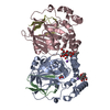


 PDBj
PDBj Assembly

