 Movie
Movie Controller


+ Open data

- Basic information
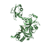
| Entry | Database: PDB / ID: 7cml | ||||||
|---|---|---|---|---|---|---|---|
| Title | The Crystal Structure of human JNK2 from Biortus. | ||||||
 Components Components | Mitogen-activated protein kinase 9 | ||||||
 Keywords Keywords |  TRANSFERASE / Protein kinase-like / transcription factor binding TRANSFERASE / Protein kinase-like / transcription factor binding | ||||||
| Function / homology |  Function and homology information Function and homology informationprotein localization to tricellular tight junction / JUN kinase activity / inflammatory response to wounding / positive regulation of macrophage derived foam cell differentiation / positive regulation of cytokine production involved in inflammatory response / positive regulation of podosome assembly / Activation of the AP-1 family of transcription factors / Fc-epsilon receptor signaling pathway / mitogen-activated protein kinase / JNK cascade ...protein localization to tricellular tight junction / JUN kinase activity / inflammatory response to wounding / positive regulation of macrophage derived foam cell differentiation / positive regulation of cytokine production involved in inflammatory response / positive regulation of podosome assembly / Activation of the AP-1 family of transcription factors / Fc-epsilon receptor signaling pathway / mitogen-activated protein kinase / JNK cascade / cellular response to cadmium ion / protein serine/threonine/tyrosine kinase activity / JNK (c-Jun kinases) phosphorylation and activation mediated by activated human TAK1 / positive regulation of protein ubiquitination / positive regulation of apoptotic signaling pathway / apoptotic signaling pathway / FCERI mediated MAPK activation / Schaffer collateral - CA1 synapse / modulation of chemical synaptic transmission / Signaling by ALK fusions and activated point mutants / regulation of circadian rhythm / cellular response to reactive oxygen species / cellular senescence / rhythmic process / positive regulation of proteasomal ubiquitin-dependent protein catabolic process / Oxidative Stress Induced Senescence / nuclear speck / protein phosphorylation / protein serine kinase activity / protein serine/threonine kinase activity / positive regulation of gene expression / mitochondrion / nucleoplasm / ATP binding / nucleus / plasma membrane / cytosol / cytoplasmSimilarity search - Function | ||||||
| Biological species |  Homo sapiens (human) Homo sapiens (human) | ||||||
| Method | X-RAY DIFFRACTION / SYNCHROTRON / MOLECULAR REPLACEMENT / Resolution: 2.15 Å | ||||||
 Authors Authors | Wang, F. / Lin, D. / Cheng, W. / Miao, Q. / Huang, Y. / Shang, H. | ||||||
 Citation Citation | Journal: To Be Published Title: The Crystal Structure of human JNK2 from Biortus. Authors: Wang, F. / Lin, D. / Cheng, W. / Miao, Q. / Huang, Y. / Shang, H. | ||||||
| History |
|
- Structure visualization
| Structure viewer | Molecule: MolmilJmol/JSmol |
|---|
- Downloads & links
-Download
| PDBx/mmCIF format | 7cml.cif.gz | 155.5 KB | Display | PDBx/mmCIF format |
|---|---|---|---|---|
| PDB format | pdb7cml.ent.gz | 116.5 KB | Display | PDB format |
| PDBx/mmJSON format | 7cml.json.gz | Tree view | PDBx/mmJSON format | |
| Others |  Other downloads Other downloads |
-Validation report
| Arichive directory | https://data.pdbj.org/pub/pdb/validation_reports/cm/7cmlftp://data.pdbj.org/pub/pdb/validation_reports/cm/7cml | HTTPS FTP |
|---|
-Related structure data
| Related structure data |  3npcS S: Starting model for refinement |
|---|---|
| Similar structure data |


-Links
 PDBj
PDBj


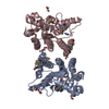
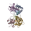
- Assembly
| Deposited unit | 
| ||||||||
|---|---|---|---|---|---|---|---|---|---|
| 1 | 
| ||||||||
| 2 | 
| ||||||||
| Unit cell |
|
-Components
| #1: Protein | Mass: 43909.484 Da / Num. of mol.: 2 / Mutation: D3K, C6V, S8N, Q14E, K203A, E204A, K250A, K251A Source method: isolated from a genetically manipulated source Source: (gene. exp.) Homo sapiens (human) / Gene: MAPK9, JNK2, PRKM9, SAPK1A / Production host:  Escherichia coli (E. coli) Escherichia coli (E. coli)References: UniProt: P45984, mitogen-activated protein kinase#2: Water | ChemComp-HOH / | Water Mass: 18.015 Da / Num. of mol.: 167 / Source method: isolated from a natural source / Formula: H2O Mass: 18.015 Da / Num. of mol.: 167 / Source method: isolated from a natural source / Formula: H2O |
|---|
-Experimental details
-Experiment
| Experiment | Method: X-RAY DIFFRACTION / Number of used crystals: 1 |
|---|
- Sample preparation
| Crystal | Density Matthews: 2.18 Å3/Da / Density % sol: 43.47 % |
|---|---|
| Crystal grow | Temperature: 293 K / Method: vapor diffusion, sitting drop / Details: 0.2M Na/K PO4, 20% PEG3,350 |
-Data collection
| Diffraction | Mean temperature: 100 K / Serial crystal experiment: N |
|---|---|
| Diffraction source | Source: SYNCHROTRON / Site: SSRF  / Beamline: BL18U1 / Wavelength: 0.97915 Å / Beamline: BL18U1 / Wavelength: 0.97915 Å |
| Detector | Type: DECTRIS PILATUS3 6M / Detector: PIXEL / Date: Apr 23, 2020 |
| Radiation | Protocol: SINGLE WAVELENGTH / Monochromatic (M) / Laue (L): M / Scattering type: x-ray |
| Radiation wavelength | Wavelength: 0.97915 Å / Relative weight: 1 |
| Reflection | Resolution: 2.15→19.616 Å / Num. obs: 39267 / % possible obs: 99.8 % / Redundancy: 9.1 % / Rmerge(I) obs: 0.141 / Net I/σ(I): 10.8 |
| Reflection shell | Resolution: 2.15→2.22 Å / Rmerge(I) obs: 1.269 / Num. unique obs: 3435 |
- Processing
| Software |
| ||||||||||||||||||||||||||||||||||||||||||||||||||||||||||||||||||||||||||||||||||||||||||||||||||||||||||||||||||||||||||||||||||||||||||||||||||||||
|---|---|---|---|---|---|---|---|---|---|---|---|---|---|---|---|---|---|---|---|---|---|---|---|---|---|---|---|---|---|---|---|---|---|---|---|---|---|---|---|---|---|---|---|---|---|---|---|---|---|---|---|---|---|---|---|---|---|---|---|---|---|---|---|---|---|---|---|---|---|---|---|---|---|---|---|---|---|---|---|---|---|---|---|---|---|---|---|---|---|---|---|---|---|---|---|---|---|---|---|---|---|---|---|---|---|---|---|---|---|---|---|---|---|---|---|---|---|---|---|---|---|---|---|---|---|---|---|---|---|---|---|---|---|---|---|---|---|---|---|---|---|---|---|---|---|---|---|---|---|---|---|
| Refinement | Method to determine structure: MOLECULAR REPLACEMENT Starting model: 3npc Resolution: 2.15→19.616 Å / Cor.coef. Fo:Fc: 0.961 / Cor.coef. Fo:Fc free: 0.938 / WRfactor Rfree: 0.327 / WRfactor Rwork: 0.284 / SU B: 7.889 / SU ML: 0.196 / Average fsc free: 0.8972 / Average fsc work: 0.9186 / Cross valid method: FREE R-VALUE / ESU R: 0.381 / ESU R Free: 0.277 Details: Hydrogens have been added in their riding positions
| ||||||||||||||||||||||||||||||||||||||||||||||||||||||||||||||||||||||||||||||||||||||||||||||||||||||||||||||||||||||||||||||||||||||||||||||||||||||
| Solvent computation | Ion probe radii: 0.8 Å / Shrinkage radii: 0.8 Å / VDW probe radii: 1.2 Å / Solvent model: MASK BULK SOLVENT | ||||||||||||||||||||||||||||||||||||||||||||||||||||||||||||||||||||||||||||||||||||||||||||||||||||||||||||||||||||||||||||||||||||||||||||||||||||||
| Displacement parameters | Biso mean: 36.292 Å2
| ||||||||||||||||||||||||||||||||||||||||||||||||||||||||||||||||||||||||||||||||||||||||||||||||||||||||||||||||||||||||||||||||||||||||||||||||||||||
| Refinement step | Cycle: LAST / Resolution: 2.15→19.616 Å
| ||||||||||||||||||||||||||||||||||||||||||||||||||||||||||||||||||||||||||||||||||||||||||||||||||||||||||||||||||||||||||||||||||||||||||||||||||||||
| Refine LS restraints |
| ||||||||||||||||||||||||||||||||||||||||||||||||||||||||||||||||||||||||||||||||||||||||||||||||||||||||||||||||||||||||||||||||||||||||||||||||||||||
| LS refinement shell |
|
