 Movie
Movie Controller

[English] 日本語
 Yorodumi
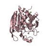
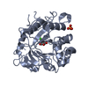
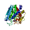
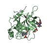
- PDB-7cjg: Structural and kinetic characterization of Porphyromonas gingival... -
+ Open data

- Basic information
| Entry | Database: PDB / ID: 7cjg | ||||||
|---|---|---|---|---|---|---|---|
| Title | Structural and kinetic characterization of Porphyromonas gingivalis glutaminyl cyclase | ||||||
 Components Components | Glutamine cyclotransferase-related protein | ||||||
 Keywords Keywords |  ANTIBIOTIC / glutaminyl cyclase / pyroglutamate / prophyromonas gingivalis ANTIBIOTIC / glutaminyl cyclase / pyroglutamate / prophyromonas gingivalis | ||||||
| Function / homology | Glutaminyl-peptide cyclotransferase-like / Peptidase M28 / Peptidase family M28 / acyltransferase activity / Prokaryotic membrane lipoprotein lipid attachment site profile. / 5,6-DIMETHYLBENZIMIDAZOLE / NITRATE ION / Glutamine cyclotransferase-related protein Function and homology information Function and homology information | ||||||
| Biological species |  Porphyromonas gingivalis (bacteria) Porphyromonas gingivalis (bacteria) | ||||||
| Method | X-RAY DIFFRACTION / MOLECULAR REPLACEMENT / Resolution: 2 Å | ||||||
 Authors Authors | Ruiz-Carrillo, D. / Lamers, S. / Feng, Q. / Yu, S. / Sun, B. / Jiang, J. / Lukman, M. | ||||||
| Funding support |  China, 1items China, 1items
| ||||||
 Citation Citation | Journal: Biol.Chem. / Year: 2021 Title: Structural and kinetic characterization of Porphyromonas gingivalis glutaminyl cyclase. Authors: Lamers, S. / Feng, Q. / Cheng, Y. / Yu, S. / Sun, B. / Lukman, M. / Jiang, J. / Ruiz-Carrillo, D. | ||||||
| History |
|
- Structure visualization
| Structure viewer | Molecule: MolmilJmol/JSmol |
|---|
- Downloads & links
-Download
| PDBx/mmCIF format | 7cjg.cif.gz | 332.5 KB | Display | PDBx/mmCIF format |
|---|---|---|---|---|
| PDB format | pdb7cjg.ent.gz | 274.8 KB | Display | PDB format |
| PDBx/mmJSON format | 7cjg.json.gz | Tree view | PDBx/mmJSON format | |
| Others |  Other downloads Other downloads |
-Validation report
| Arichive directory | https://data.pdbj.org/pub/pdb/validation_reports/cj/7cjgftp://data.pdbj.org/pub/pdb/validation_reports/cj/7cjg | HTTPS FTP |
|---|
-Related structure data
| Related structure data |  7cjeSC S: Starting model for refinement C: citing same article ( |
|---|---|
| Similar structure data |


-Links
 PDBj
PDBj


- Assembly
| Deposited unit | 
| ||||||||||||||||||||||||
|---|---|---|---|---|---|---|---|---|---|---|---|---|---|---|---|---|---|---|---|---|---|---|---|---|---|
| 1 | 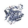
| ||||||||||||||||||||||||
| 2 | 
| ||||||||||||||||||||||||
| Unit cell |
| ||||||||||||||||||||||||
| Noncrystallographic symmetry (NCS) | NCS domain:
NCS domain segments:
|
-Components
-Protein , 1 types, 2 molecules AB
| #1: Protein | Mass: 36565.383 Da / Num. of mol.: 2 Source method: isolated from a genetically manipulated source Source: (gene. exp.) Porphyromonas gingivalis (strain ATCC BAA-308 / W83) (bacteria)Strain: ATCC BAA-308 / W83 / Gene: PG_2157 / Production host: Escherichia coli BL21(DE3) (bacteria) / References: UniProt: Q7MT37 |
|---|
-Non-polymers , 6 types, 344 molecules 


| #2: Chemical | Dimethylbenzimidazole Mass: 146.189 Da / Num. of mol.: 2 / Source method: obtained synthetically / Formula: C9H10N2 Mass: 146.189 Da / Num. of mol.: 2 / Source method: obtained synthetically / Formula: C9H10N2#3: Chemical | Glycerol Mass: 92.094 Da / Num. of mol.: 2 / Source method: obtained synthetically / Formula: C3H8O3 Mass: 92.094 Da / Num. of mol.: 2 / Source method: obtained synthetically / Formula: C3H8O3#4: Chemical | Nitrate Mass: 62.005 Da / Num. of mol.: 3 / Source method: obtained synthetically / Formula: NO3 Mass: 62.005 Da / Num. of mol.: 3 / Source method: obtained synthetically / Formula: NO3#5: Chemical |  Mass: 24.305 Da / Num. of mol.: 2 / Source method: obtained synthetically / Formula: Mg Mass: 24.305 Da / Num. of mol.: 2 / Source method: obtained synthetically / Formula: Mg#6: Chemical |  Mass: 65.409 Da / Num. of mol.: 2 / Source method: obtained synthetically / Formula: Zn Mass: 65.409 Da / Num. of mol.: 2 / Source method: obtained synthetically / Formula: Zn#7: Water | ChemComp-HOH / | WaterMass: 18.015 Da / Num. of mol.: 333 / Source method: isolated from a natural source / Formula: H2O |
|---|
-Details
| Has ligand of interest | Y |
|---|
-Experimental details
-Experiment
| Experiment | Method: X-RAY DIFFRACTION / Number of used crystals: 1 |
|---|
- Sample preparation
| Crystal | Density Matthews: 2.7 Å3/Da / Density % sol: 54.52 % |
|---|---|
| Crystal grow | Temperature: 287.15 K / Method: vapor diffusion, hanging drop / pH: 8 Details: Purified PgQC was concentrated up to 50 mg/ml and it was mixed in a 1:0.7 volume ratio with the crystallization buffer 0.1 M TRIS pH 8.0, 0.3 M MgNO3(H2O)6, 24% (w/v) PEG8000) incubating at 14 C |
-Data collection
| Diffraction | Mean temperature: 150 K / Serial crystal experiment: N |
|---|---|
| Diffraction source | Source: SEALED TUBE / Type: BRUKER IMUS MICROFOCUS / Wavelength: 1.54184 Å |
| Detector | Type: Bruker PHOTON II / Detector: PIXEL / Date: Jun 22, 2020 |
| Radiation | Protocol: SINGLE WAVELENGTH / Monochromatic (M) / Laue (L): M / Scattering type: x-ray |
| Radiation wavelength | Wavelength: 1.54184 Å / Relative weight: 1 |
| Reflection | Resolution: 2→45.56 Å / Num. obs: 103509 / % possible obs: 99.9 % / Redundancy: 24 % / Biso Wilson estimate: 31.75 Å2 / Rmerge(I) obs: 0.2057 / Net I/σ(I): 27.83 |
| Reflection shell | Resolution: 2→2.07 Å / Redundancy: 15.1 % / Rmerge(I) obs: 1.565 / Mean I/σ(I) obs: 1.76 / Num. unique obs: 5383 / % possible all: 100 |
- Processing
| Software |
| |||||||||||||||||||||||||||||||||||||||||||||||||||||||||||||||||||||||||||||||||||||||||||||||||||||||||||||||||||||||||||||||||||||||||||||||||||||||||||||||||||||||||||||||||||||||||||||||||||||||||||||||||||||||||||||||||||||||||||||||||||||||||||||||||||||||||||||||||||
|---|---|---|---|---|---|---|---|---|---|---|---|---|---|---|---|---|---|---|---|---|---|---|---|---|---|---|---|---|---|---|---|---|---|---|---|---|---|---|---|---|---|---|---|---|---|---|---|---|---|---|---|---|---|---|---|---|---|---|---|---|---|---|---|---|---|---|---|---|---|---|---|---|---|---|---|---|---|---|---|---|---|---|---|---|---|---|---|---|---|---|---|---|---|---|---|---|---|---|---|---|---|---|---|---|---|---|---|---|---|---|---|---|---|---|---|---|---|---|---|---|---|---|---|---|---|---|---|---|---|---|---|---|---|---|---|---|---|---|---|---|---|---|---|---|---|---|---|---|---|---|---|---|---|---|---|---|---|---|---|---|---|---|---|---|---|---|---|---|---|---|---|---|---|---|---|---|---|---|---|---|---|---|---|---|---|---|---|---|---|---|---|---|---|---|---|---|---|---|---|---|---|---|---|---|---|---|---|---|---|---|---|---|---|---|---|---|---|---|---|---|---|---|---|---|---|---|---|---|---|---|---|---|---|---|---|---|---|---|---|---|---|---|---|---|---|---|---|---|---|---|---|---|---|---|---|---|---|---|---|---|---|---|---|---|---|---|---|---|---|---|---|---|---|---|---|---|
| Refinement | Method to determine structure: MOLECULAR REPLACEMENT Starting model: 7CJE Resolution: 2→32.06 Å / SU ML: 0.258 / Cross valid method: THROUGHOUT / σ(F): 1.358 / Phase error: 21.987 Stereochemistry target values: GEOSTD + MONOMER LIBRARY + CDL V1.2
| |||||||||||||||||||||||||||||||||||||||||||||||||||||||||||||||||||||||||||||||||||||||||||||||||||||||||||||||||||||||||||||||||||||||||||||||||||||||||||||||||||||||||||||||||||||||||||||||||||||||||||||||||||||||||||||||||||||||||||||||||||||||||||||||||||||||||||||||||||
| Solvent computation | Shrinkage radii: 0.9 Å / VDW probe radii: 1.11 Å / Solvent model: FLAT BULK SOLVENT MODEL | |||||||||||||||||||||||||||||||||||||||||||||||||||||||||||||||||||||||||||||||||||||||||||||||||||||||||||||||||||||||||||||||||||||||||||||||||||||||||||||||||||||||||||||||||||||||||||||||||||||||||||||||||||||||||||||||||||||||||||||||||||||||||||||||||||||||||||||||||||
| Displacement parameters | Biso mean: 48.83 Å2 | |||||||||||||||||||||||||||||||||||||||||||||||||||||||||||||||||||||||||||||||||||||||||||||||||||||||||||||||||||||||||||||||||||||||||||||||||||||||||||||||||||||||||||||||||||||||||||||||||||||||||||||||||||||||||||||||||||||||||||||||||||||||||||||||||||||||||||||||||||
| Refinement step | Cycle: LAST / Resolution: 2→32.06 Å
| |||||||||||||||||||||||||||||||||||||||||||||||||||||||||||||||||||||||||||||||||||||||||||||||||||||||||||||||||||||||||||||||||||||||||||||||||||||||||||||||||||||||||||||||||||||||||||||||||||||||||||||||||||||||||||||||||||||||||||||||||||||||||||||||||||||||||||||||||||
| Refine LS restraints |
| |||||||||||||||||||||||||||||||||||||||||||||||||||||||||||||||||||||||||||||||||||||||||||||||||||||||||||||||||||||||||||||||||||||||||||||||||||||||||||||||||||||||||||||||||||||||||||||||||||||||||||||||||||||||||||||||||||||||||||||||||||||||||||||||||||||||||||||||||||
| Refine LS restraints NCS |
| |||||||||||||||||||||||||||||||||||||||||||||||||||||||||||||||||||||||||||||||||||||||||||||||||||||||||||||||||||||||||||||||||||||||||||||||||||||||||||||||||||||||||||||||||||||||||||||||||||||||||||||||||||||||||||||||||||||||||||||||||||||||||||||||||||||||||||||||||||
| LS refinement shell |
| |||||||||||||||||||||||||||||||||||||||||||||||||||||||||||||||||||||||||||||||||||||||||||||||||||||||||||||||||||||||||||||||||||||||||||||||||||||||||||||||||||||||||||||||||||||||||||||||||||||||||||||||||||||||||||||||||||||||||||||||||||||||||||||||||||||||||||||||||||
| Refinement TLS params. | Method: refined / Refine-ID: X-RAY DIFFRACTION
| |||||||||||||||||||||||||||||||||||||||||||||||||||||||||||||||||||||||||||||||||||||||||||||||||||||||||||||||||||||||||||||||||||||||||||||||||||||||||||||||||||||||||||||||||||||||||||||||||||||||||||||||||||||||||||||||||||||||||||||||||||||||||||||||||||||||||||||||||||
| Refinement TLS group |
|
