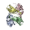
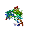
 Movie
Movie Controller

[English] 日本語
 Yorodumi
- PDB-7cgq: Crystal structure of Azospirillum brasilense L-arabinose 1-dehydr... -
+ Open data

- Basic information
| Entry | Database: PDB / ID: 7cgq | ||||||
|---|---|---|---|---|---|---|---|
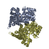
| Title | Crystal structure of Azospirillum brasilense L-arabinose 1-dehydrogenase E147A mutant (NADP and L-arabinose bound form) | ||||||
 Components Components | L-arabinose 1-dehydrogenase (NAD(P)(+)) | ||||||
 Keywords Keywords |  OXIDOREDUCTASE / L-ARABINOSE METABOLISM / NADP-DEPENDENT DEHYDROGENASE / GFO/IDH/MOCA PROTEIN FAMILY OXIDOREDUCTASE / L-ARABINOSE METABOLISM / NADP-DEPENDENT DEHYDROGENASE / GFO/IDH/MOCA PROTEIN FAMILY | ||||||
| Function / homology |  Function and homology information Function and homology informationL-arabinose 1-dehydrogenase [NAD(P)+] / L-arabinose catabolic process / L-arabinose 1-dehydrogenase (NADP+) activity / L-arabinose 1-dehydrogenase (NAD+) activity / galactose 1-dehydrogenase (NADP+) / galactose 1-dehydrogenase (NADP+) activity / D-galactose 1-dehydrogenase / galactose 1-dehydrogenase activity / L-arabinose catabolic process to 2-oxoglutarate / oxidoreductase activity, acting on the CH-OH group of donors, NAD or NADP as acceptor ...L-arabinose 1-dehydrogenase [NAD(P)+] / L-arabinose catabolic process / L-arabinose 1-dehydrogenase (NADP+) activity / L-arabinose 1-dehydrogenase (NAD+) activity / galactose 1-dehydrogenase (NADP+) / galactose 1-dehydrogenase (NADP+) activity / D-galactose 1-dehydrogenase / galactose 1-dehydrogenase activity / L-arabinose catabolic process to 2-oxoglutarate / oxidoreductase activity, acting on the CH-OH group of donors, NAD or NADP as acceptor / NADP+ binding / NAD+ bindingSimilarity search - Function | ||||||
| Biological species |  Azospirillum brasilense (bacteria) Azospirillum brasilense (bacteria) | ||||||
| Method | X-RAY DIFFRACTION / SYNCHROTRON / MOLECULAR REPLACEMENT / Resolution: 2.208 Å | ||||||
 Authors Authors | Yoshiwara, K. / Watanabe, Y. / Watanabe, S. | ||||||
 Citation Citation | Journal: Biochem.Biophys.Res.Commun. / Year: 2020 Title: Crystal structure of bacterial L-arabinose 1-dehydrogenase in complex with L-arabinose and NADP+ Authors: Yoshiwara, K. / Watanabe, S. / Watanabe, Y. | ||||||
| History |
|
- Structure visualization
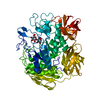
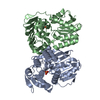
| Structure viewer | Molecule: MolmilJmol/JSmol |
|---|
- Downloads & links
-Download
| PDBx/mmCIF format | 7cgq.cif.gz | 252.3 KB | Display | PDBx/mmCIF format |
|---|---|---|---|---|
| PDB format | pdb7cgq.ent.gz | 202.2 KB | Display | PDB format |
| PDBx/mmJSON format | 7cgq.json.gz | Tree view | PDBx/mmJSON format | |
| Others |  Other downloads Other downloads |
-Validation report
| Arichive directory | https://data.pdbj.org/pub/pdb/validation_reports/cg/7cgqftp://data.pdbj.org/pub/pdb/validation_reports/cg/7cgq | HTTPS FTP |
|---|
-Related structure data
| Related structure data | 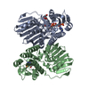 7cgrC 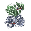 6jnkS S: Starting model for refinement C: citing same article ( |
|---|---|
| Similar structure data |


-Links
 PDBj
PDBj- Assembly
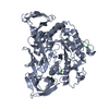
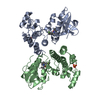
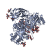
| Deposited unit | 
| ||||||||
|---|---|---|---|---|---|---|---|---|---|
| 1 | 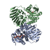
| ||||||||
| 2 | 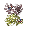
| ||||||||
| Unit cell |
|
-Components
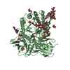
| #1: Protein | Mass: 34967.727 Da / Num. of mol.: 4 / Mutation: E147A Source method: isolated from a genetically manipulated source Source: (gene. exp.) Azospirillum brasilense (bacteria) / Gene: araA / Production host: Escherichia coli (E. coli)References: UniProt: Q53TZ2, L-arabinose 1-dehydrogenase [NAD(P)+], galactose 1-dehydrogenase (NADP+), D-galactose 1-dehydrogenase#2: Chemical | ChemComp-NAP / Nicotinamide adenine dinucleotide phosphate  Mass: 743.405 Da / Num. of mol.: 4 / Source method: obtained synthetically / Formula: C21H28N7O17P3 / Feature type: SUBJECT OF INVESTIGATION Mass: 743.405 Da / Num. of mol.: 4 / Source method: obtained synthetically / Formula: C21H28N7O17P3 / Feature type: SUBJECT OF INVESTIGATION#3: Sugar | ChemComp-ARA / | Arabinose  Type: L-saccharide, alpha linking / Mass: 150.130 Da / Num. of mol.: 1 Type: L-saccharide, alpha linking / Mass: 150.130 Da / Num. of mol.: 1Source method: isolated from a genetically manipulated source Formula: C5H10O5 #4: Water | ChemComp-HOH / | Water Mass: 18.015 Da / Num. of mol.: 342 / Source method: isolated from a natural source / Formula: H2O Mass: 18.015 Da / Num. of mol.: 342 / Source method: isolated from a natural source / Formula: H2OHas ligand of interest | Y | |
|---|
-Experimental details
-Experiment
| Experiment | Method: X-RAY DIFFRACTION / Number of used crystals: 1 |
|---|
- Sample preparation
| Crystal | Density Matthews: 2.61 Å3/Da / Density % sol: 52.91 % Description: The entry contains friedel pairs in F_plus/minus columns and I_plus/minus columns |
|---|---|
| Crystal grow | Temperature: 293 K / Method: vapor diffusion, sitting drop / pH: 7 Details: 0.2M Ammonium citrate tribasic pH 7.0, 18.5% PEG 3350 |
-Data collection
| Diffraction | Mean temperature: 100 K / Serial crystal experiment: N |
|---|---|
| Diffraction source | Source: SYNCHROTRON / Site: SPring-8  / Beamline: BL41XU / Wavelength: 1 Å / Beamline: BL41XU / Wavelength: 1 Å |
| Detector | Type: DECTRIS EIGER X 16M / Detector: PIXEL / Date: Feb 16, 2020 |
| Radiation | Protocol: SINGLE WAVELENGTH / Monochromatic (M) / Laue (L): M / Scattering type: x-ray |
| Radiation wavelength | Wavelength: 1 Å / Relative weight: 1 |
| Reflection | Resolution: 2.208→50 Å / Num. obs: 82746 / % possible obs: 95.7 % / Redundancy: 1.8 % / CC1/2: 0.996 / Rmerge(I) obs: 0.085 / Rrim(I) all: 0.084 / Net I/σ(I): 6.45 |
| Reflection shell | Resolution: 2.21→2.34 Å / Rmerge(I) obs: 0.74 / Mean I/σ(I) obs: 1 / Num. unique obs: 21839 / CC1/2: 0.664 / Rrim(I) all: 0.798 |
- Processing
| Software |
| ||||||||||||||||||||||||||||||||||||||||||||||||||||||||||||||||||||||||||||||||||||||||||||||||||||||||||||||||||||||||||||||||||||||||||||||||||||||||||||||||||
|---|---|---|---|---|---|---|---|---|---|---|---|---|---|---|---|---|---|---|---|---|---|---|---|---|---|---|---|---|---|---|---|---|---|---|---|---|---|---|---|---|---|---|---|---|---|---|---|---|---|---|---|---|---|---|---|---|---|---|---|---|---|---|---|---|---|---|---|---|---|---|---|---|---|---|---|---|---|---|---|---|---|---|---|---|---|---|---|---|---|---|---|---|---|---|---|---|---|---|---|---|---|---|---|---|---|---|---|---|---|---|---|---|---|---|---|---|---|---|---|---|---|---|---|---|---|---|---|---|---|---|---|---|---|---|---|---|---|---|---|---|---|---|---|---|---|---|---|---|---|---|---|---|---|---|---|---|---|---|---|---|---|---|---|
| Refinement | Method to determine structure: MOLECULAR REPLACEMENT Starting model: 6JNK Resolution: 2.208→49.385 Å / SU ML: 0.23 / Cross valid method: THROUGHOUT / σ(F): 1.34 / Phase error: 24.2 / Stereochemistry target values: ML Details: The entry contains friedel pairs in F_plus/minus columns and I_plus/minus columns
| ||||||||||||||||||||||||||||||||||||||||||||||||||||||||||||||||||||||||||||||||||||||||||||||||||||||||||||||||||||||||||||||||||||||||||||||||||||||||||||||||||
| Solvent computation | Shrinkage radii: 0.9 Å / VDW probe radii: 1.11 Å / Solvent model: FLAT BULK SOLVENT MODEL | ||||||||||||||||||||||||||||||||||||||||||||||||||||||||||||||||||||||||||||||||||||||||||||||||||||||||||||||||||||||||||||||||||||||||||||||||||||||||||||||||||
| Displacement parameters | Biso max: 93.98 Å2 / Biso mean: 32.0001 Å2 / Biso min: 14.51 Å2 | ||||||||||||||||||||||||||||||||||||||||||||||||||||||||||||||||||||||||||||||||||||||||||||||||||||||||||||||||||||||||||||||||||||||||||||||||||||||||||||||||||
| Refinement step | Cycle: final / Resolution: 2.208→49.385 Å
| ||||||||||||||||||||||||||||||||||||||||||||||||||||||||||||||||||||||||||||||||||||||||||||||||||||||||||||||||||||||||||||||||||||||||||||||||||||||||||||||||||
| LS refinement shell | Refine-ID: X-RAY DIFFRACTION / Rfactor Rfree error: 0
|
