 Movie
Movie Controller


+ Open data

- Basic information
| Entry | Database: PDB / ID: 7c9c | ||||||||||||
|---|---|---|---|---|---|---|---|---|---|---|---|---|---|
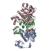
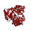
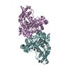
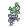
| Title | Human DMC1 pre-synaptic complexes | ||||||||||||
 Components Components |
| ||||||||||||
 Keywords Keywords | RECOMBINATION/DNA / meiotic homologous recombination /  DNA repair / ATPase / Recombination / RECOMBINATION-DNA complex DNA repair / ATPase / Recombination / RECOMBINATION-DNA complex | ||||||||||||
| Function / homology |  Function and homology information Function and homology informationfemale gamete generation / chromosome organization involved in meiotic cell cycle / DNA recombinase assembly / homologous chromosome pairing at meiosis / double-strand break repair involved in meiotic recombination / mitotic recombination / DNA strand invasion / DNA strand exchange activity / lateral element / oocyte maturation ...female gamete generation / chromosome organization involved in meiotic cell cycle / DNA recombinase assembly / homologous chromosome pairing at meiosis / double-strand break repair involved in meiotic recombination / mitotic recombination / DNA strand invasion / DNA strand exchange activity / lateral element / oocyte maturation / reciprocal meiotic recombination / ATP-dependent DNA damage sensor activity / male meiosis I / spermatid development / ATP-dependent activity, acting on DNA / ovarian follicle development / meiotic cell cycle / condensed nuclear chromosome / Meiotic recombination / single-stranded DNA binding / site of double-strand break / chromosome / double-stranded DNA binding / spermatogenesis / chromosome, telomeric region / ATP hydrolysis activity / DNA binding / ATP binding / identical protein binding / nucleusSimilarity search - Function | ||||||||||||
| Biological species |  Homo sapiens (human) Homo sapiens (human)synthetic construct (others) | ||||||||||||
| Method | ELECTRON MICROSCOPY / helical reconstruction / cryo EM / Resolution: 3.33 Å | ||||||||||||
 Authors Authors | Luo, S.C. / Yeh, H.Y. / Chi, P. / Ho, M.C. / Tsai, M.D. | ||||||||||||
| Funding support |  Taiwan, 3items Taiwan, 3items
| ||||||||||||
 Citation Citation | Journal: Nat Commun / Year: 2021 Title: Identification of fidelity-governing factors in human recombinases DMC1 and RAD51 from cryo-EM structures. Authors: Shih-Chi Luo / Hsin-Yi Yeh / Wei-Hsuan Lan / Yi-Min Wu / Cheng-Han Yang / Hao-Yen Chang / Guan-Chin Su / Chia-Yi Lee / Wen-Jin Wu / Hung-Wen Li / Meng-Chiao Ho / Peter Chi / Ming-Daw Tsai / Abstract: Both high-fidelity and mismatch-tolerant recombination, catalyzed by RAD51 and DMC1 recombinases, respectively, are indispensable for genomic integrity. Here, we use cryo-EM, MD simulation and ...Both high-fidelity and mismatch-tolerant recombination, catalyzed by RAD51 and DMC1 recombinases, respectively, are indispensable for genomic integrity. Here, we use cryo-EM, MD simulation and functional analysis to elucidate the structural basis for the mismatch tolerance of DMC1. Structural analysis of DMC1 presynaptic and postsynaptic complexes suggested that the lineage-specific Loop 1 Gln244 (Met243 in RAD51) may help stabilize DNA backbone, whereas Loop 2 Pro274 and Gly275 (Val273/Asp274 in RAD51) may provide an open "triplet gate" for mismatch tolerance. In support, DMC1-Q244M displayed marked increase in DNA dynamics, leading to unobservable DNA map. MD simulation showed highly dispersive mismatched DNA ensemble in RAD51 but well-converged DNA in DMC1 and RAD51-V273P/D274G. Replacing Loop 1 or Loop 2 residues in DMC1 with RAD51 counterparts enhanced DMC1 fidelity, while reciprocal mutations in RAD51 attenuated its fidelity. Our results show that three Loop 1/Loop 2 residues jointly enact contrasting fidelities of DNA recombinases. | ||||||||||||
| History |
|
- Structure visualization
| Movie |
Movie viewer |
|---|---|
| Structure viewer | Molecule: MolmilJmol/JSmol |
- Downloads & links
-Download
| PDBx/mmCIF format | 7c9c.cif.gz | 167.9 KB | Display | PDBx/mmCIF format |
|---|---|---|---|---|
| PDB format | pdb7c9c.ent.gz | 138.9 KB | Display | PDB format |
| PDBx/mmJSON format | 7c9c.json.gz | Tree view | PDBx/mmJSON format | |
| Others |  Other downloads Other downloads |
-Validation report
| Arichive directory | https://data.pdbj.org/pub/pdb/validation_reports/c9/7c9cftp://data.pdbj.org/pub/pdb/validation_reports/c9/7c9c | HTTPS FTP |
|---|
-Related structure data
| Related structure data |  30311MC 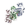 7c98C 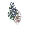 7c99C 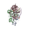 7c9aC  7cgyC M: map data used to model this data C: citing same article ( |
|---|---|
| Similar structure data |


-Links
 PDBj
PDBj


- Assembly
| Deposited unit | 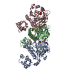
|
|---|---|
| 1 |
|
-Components
| #1: DNA chain | Mass: 2692.778 Da / Num. of mol.: 1 / Source method: obtained synthetically / Source: (synth.) synthetic construct (others) | ||||||||
|---|---|---|---|---|---|---|---|---|---|
| #2: Protein | Genetic recombination Mass: 37731.031 Da / Num. of mol.: 3 Source method: isolated from a genetically manipulated source Source: (gene. exp.) Homo sapiens (human) / Gene: DMC1, DMC1H, LIM15 / Production host:  Escherichia coli (E. coli) / Strain (production host): BLR / References: UniProt: Q14565 Escherichia coli (E. coli) / Strain (production host): BLR / References: UniProt: Q14565#3: Chemical |   Mass: 40.078 Da / Num. of mol.: 3 / Source method: obtained synthetically / Formula: Ca Mass: 40.078 Da / Num. of mol.: 3 / Source method: obtained synthetically / Formula: Ca#4: Chemical |   Mass: 506.196 Da / Num. of mol.: 3 / Source method: obtained synthetically / Formula: C10H17N6O12P3 / Comment: AMP-PNP, energy-carrying molecule analogue*YM Mass: 506.196 Da / Num. of mol.: 3 / Source method: obtained synthetically / Formula: C10H17N6O12P3 / Comment: AMP-PNP, energy-carrying molecule analogue*YMHas ligand of interest | N | Sequence details | Authors know the sequence of chains D: ...Authors know the sequence of chains D: TTATGTTCAT | |
-Experimental details
-Experiment
| Experiment | Method: ELECTRON MICROSCOPY |
|---|---|
| EM experiment | Aggregation state: FILAMENT / 3D reconstruction method: helical reconstruction |
- Sample preparation
| Component | Name: DMC1-ssDNA filament / Type: COMPLEX / Entity ID: #1-#2 / Source: RECOMBINANT |
|---|---|
| Source (natural) | Organism: Homo sapiens (human) |
| Source (recombinant) | Organism: Escherichia coli (E. coli) |
| Buffer solution | pH: 7.5 Details: 25 mM Tris-HCl, pH 7.5, 50 mM KCl and 1 mM dithiothreitol) containing 2 mM AMP-PNP and 5 mM CaCl2 |
| Specimen | Embedding applied: NO / Shadowing applied: NO / Staining applied: NO / Vitrification applied: YES Details: protein sample were applied onto a pre-glow-discharged graphene-oxide coated Quantifoil holey carbon grid (1.2/1.3, 200 mesh) using published protocol |
| Specimen support | Grid material: GRAPHENE OXIDE / Grid mesh size: 200 divisions/in. / Grid type: Quantifoil R1.2/1.3 |
| Vitrification | Instrument: FEI VITROBOT MARK IV / Cryogen name: ETHANE / Humidity: 100 % / Chamber temperature: 295 K Details: The grids were blotted for 1 sec at 22 degree C with 100% relative humidity and plunge-frozen in liquid ethane cooled by liquid nitrogen using a Vitrobot Mark IV (Thermo Fisher). |
- Electron microscopy imaging
| Experimental equipment |  Model: Titan Krios / Image courtesy: FEI Company |
|---|---|
| Microscopy | Model: FEI TITAN KRIOS |
| Electron gun | Electron source: FIELD EMISSION GUN / Accelerating voltage: 300 kV / Illumination mode: FLOOD BEAM |
| Electron lens | Mode: BRIGHT FIELDBright-field microscopy / Nominal magnification: 165000 X / Nominal defocus max: 2500 nm / Nominal defocus min: 1500 nm / Cs: 2.7 mm / C2 aperture diameter: 70 µm / Alignment procedure: COMA FREE |
| Specimen holder | Cryogen: NITROGEN / Specimen holder model: FEI TITAN KRIOS AUTOGRID HOLDER / Residual tilt: 10 mradians |
| Image recording | Average exposure time: 5 sec. / Electron dose: 50 e/Å2 / Detector mode: COUNTING / Film or detector model: GATAN K2 QUANTUM (4k x 4k) / Num. of grids imaged: 1 |
| EM imaging optics | Energyfilter name: GIF Quantum LS / Energyfilter slit width: 30 eV |
| Image scans | Sampling size: 5 µm / Width: 3838 / Height: 3710 / Movie frames/image: 50 / Used frames/image: 1-50 |
- Processing
| EM software |
| ||||||||||||||||||||||||
|---|---|---|---|---|---|---|---|---|---|---|---|---|---|---|---|---|---|---|---|---|---|---|---|---|---|
| CTF correction | Type: PHASE FLIPPING AND AMPLITUDE CORRECTION | ||||||||||||||||||||||||
| Helical symmerty | Angular rotation/subunit: 55.48 ° / Axial rise/subunit: 15.71 Å / Axial symmetry: C1 | ||||||||||||||||||||||||
| Particle selection | Num. of particles selected: 192466 | ||||||||||||||||||||||||
| 3D reconstruction | Resolution: 3.33 Å / Resolution method: FSC 0.143 CUT-OFF / Num. of particles: 71790 / Symmetry type: HELICAL |
