 Movie
Movie Controller


+ Open data

- Basic information
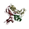
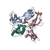
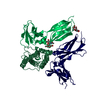
| Entry | Database: PDB / ID: 7c3m | ||||||
|---|---|---|---|---|---|---|---|
| Title | Structure of FERM protein | ||||||
 Components Components | Fermitin family homolog 3,Fermitin family homolog 3,Fermitin family homolog 3 | ||||||
 Keywords Keywords |  SIGNALING PROTEIN / FERM Protein / Kindlin / Integrin SIGNALING PROTEIN / FERM Protein / Kindlin / Integrin | ||||||
| Function / homology |  Function and homology information Function and homology informationcell-substrate junction / regulation of cell-cell adhesion mediated by integrin / integrin activation / podosome / leukocyte cell-cell adhesion / substrate adhesion-dependent cell spreading / cell-matrix adhesion / platelet alpha granule lumen / cell projection / integrin-mediated signaling pathway ...cell-substrate junction / regulation of cell-cell adhesion mediated by integrin / integrin activation / podosome / leukocyte cell-cell adhesion / substrate adhesion-dependent cell spreading / cell-matrix adhesion / platelet alpha granule lumen / cell projection / integrin-mediated signaling pathway / platelet aggregation / integrin binding / Platelet degranulation / positive regulation of cell migration / lipid binding / extracellular exosome / extracellular region / membraneSimilarity search - Function | ||||||
| Biological species |  Homo sapiens (human) Homo sapiens (human) | ||||||
| Method | X-RAY DIFFRACTION / SYNCHROTRON / SAD / Resolution: 3.6 Å | ||||||
 Authors Authors | Bu, W. / Loh, Z.Y. / Jin, S. / Basu, S. / Ero, R. / Park, J.E. / Yan, X. / Wang, M. / Sze, S.K. / Tan, S.M. / Gao, Y.G. | ||||||
| Funding support |  Singapore, 1items Singapore, 1items
| ||||||
 Citation Citation | Journal: Plos Biol. / Year: 2020 Title: Structural basis of human full-length kindlin-3 homotrimer in an auto-inhibited state. Authors: Bu, W. / Levitskaya, Z. / Loh, Z.Y. / Jin, S. / Basu, S. / Ero, R. / Yan, X. / Wang, M. / Ngan, S.F.C. / Sze, S.K. / Tan, S.M. / Gao, Y.G. | ||||||
| History |
|
- Structure visualization
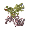
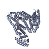
| Structure viewer | Molecule: MolmilJmol/JSmol |
|---|
- Downloads & links
-Download
| PDBx/mmCIF format | 7c3m.cif.gz | 670.1 KB | Display | PDBx/mmCIF format |
|---|---|---|---|---|
| PDB format | pdb7c3m.ent.gz | 564.8 KB | Display | PDB format |
| PDBx/mmJSON format | 7c3m.json.gz | Tree view | PDBx/mmJSON format | |
| Others |  Other downloads Other downloads |
-Validation report
| Arichive directory | https://data.pdbj.org/pub/pdb/validation_reports/c3/7c3mftp://data.pdbj.org/pub/pdb/validation_reports/c3/7c3m | HTTPS FTP |
|---|
-Related structure data
| Similar structure data |
|---|


-Links
 PDBj
PDBj

- Assembly
| Deposited unit | 
| ||||||||
|---|---|---|---|---|---|---|---|---|---|
| 1 | 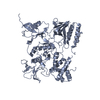
| ||||||||
| 2 | 
| ||||||||
| 3 | 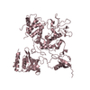
| ||||||||
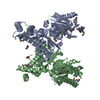
| Unit cell |
|
-Components
| #1: Protein | Mass: 76462.750 Da / Num. of mol.: 3 Source method: isolated from a genetically manipulated source Source: (gene. exp.) Homo sapiens (human) / Gene: FERMT3, KIND3, MIG2B, URP2 / Production host:  Escherichia coli (E. coli) / References: UniProt: Q86UX7 Escherichia coli (E. coli) / References: UniProt: Q86UX7Sequence details | The authors know the sequence of Residues 309-349, EVGEPAGTDPGLDDLDVALSNLEVKLEGSAPTDVLDSLTTI but ...The authors know the sequence of Residues 309-349, EVGEPAGTDP | |
|---|
-Experimental details
-Experiment
| Experiment | Method: X-RAY DIFFRACTION / Number of used crystals: 1 |
|---|
- Sample preparation
| Crystal | Density Matthews: 3.62 Å3/Da / Density % sol: 66.02 % |
|---|---|
| Crystal grow | Temperature: 286 K / Method: vapor diffusion, hanging drop Details: 3M sodium formate pH 7.0, 3% w/v xylitol, and 9% glycerol |
-Data collection
| Diffraction | Mean temperature: 100 K / Serial crystal experiment: N |
|---|---|
| Diffraction source | Source: SYNCHROTRON / Site: SLS  / Beamline: X06DA / Wavelength: 1 Å / Beamline: X06DA / Wavelength: 1 Å |
| Detector | Type: DECTRIS PILATUS 2M-F / Detector: PIXEL / Date: Sep 23, 2017 |
| Radiation | Protocol: SINGLE WAVELENGTH / Monochromatic (M) / Laue (L): M / Scattering type: x-ray |
| Radiation wavelength | Wavelength: 1 Å / Relative weight: 1 |
| Reflection | Resolution: 3.6→50 Å / Num. obs: 37501 / % possible obs: 99.9 % / Redundancy: 22.4 % / CC1/2: 0.999 / Net I/σ(I): 14.7 |
| Reflection shell | Resolution: 3.6→3.69 Å / Redundancy: 23.5 % / Mean I/σ(I) obs: 0.97 / Num. unique obs: 3704 / CC1/2: 0.547 / % possible all: 100 |
- Processing
| Software |
| ||||||||||||||||||||||||||||||||||||||||||||||||||||||||||||||||||||||||||||||||||||||||||||||||||
|---|---|---|---|---|---|---|---|---|---|---|---|---|---|---|---|---|---|---|---|---|---|---|---|---|---|---|---|---|---|---|---|---|---|---|---|---|---|---|---|---|---|---|---|---|---|---|---|---|---|---|---|---|---|---|---|---|---|---|---|---|---|---|---|---|---|---|---|---|---|---|---|---|---|---|---|---|---|---|---|---|---|---|---|---|---|---|---|---|---|---|---|---|---|---|---|---|---|---|---|
| Refinement | Method to determine structure: SAD / Resolution: 3.6→49.202 Å / SU ML: 0.55 / Cross valid method: FREE R-VALUE / σ(F): 1.34 / Phase error: 34.34 / Stereochemistry target values: ML
| ||||||||||||||||||||||||||||||||||||||||||||||||||||||||||||||||||||||||||||||||||||||||||||||||||
| Solvent computation | Shrinkage radii: 0.9 Å / VDW probe radii: 1.11 Å / Solvent model: FLAT BULK SOLVENT MODEL | ||||||||||||||||||||||||||||||||||||||||||||||||||||||||||||||||||||||||||||||||||||||||||||||||||
| Refinement step | Cycle: LAST / Resolution: 3.6→49.202 Å
| ||||||||||||||||||||||||||||||||||||||||||||||||||||||||||||||||||||||||||||||||||||||||||||||||||
| Refine LS restraints |
| ||||||||||||||||||||||||||||||||||||||||||||||||||||||||||||||||||||||||||||||||||||||||||||||||||
| LS refinement shell |
| ||||||||||||||||||||||||||||||||||||||||||||||||||||||||||||||||||||||||||||||||||||||||||||||||||
| Refinement TLS params. | Method: refined / Origin x: -17.2247 Å / Origin y: 204.2496 Å / Origin z: 306.9721 Å
| ||||||||||||||||||||||||||||||||||||||||||||||||||||||||||||||||||||||||||||||||||||||||||||||||||
| Refinement TLS group | Selection details: all |
