 Movie
Movie Controller

[English] 日本語
 Yorodumi
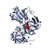
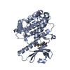
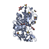
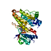
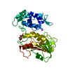
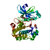
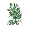
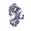
- PDB-7c3b: Ferredoxin reductase in carbazole 1,9a-dioxygenase (FAD apo form) -
+ Open data

- Basic information
| Entry | Database: PDB / ID: 7c3b | ||||||
|---|---|---|---|---|---|---|---|
| Title | Ferredoxin reductase in carbazole 1,9a-dioxygenase (FAD apo form) | ||||||
 Components Components | Ferredoxin reductase component of carbazole | ||||||
 Keywords Keywords |  OXIDOREDUCTASE / Rieske non-heme iron oxygenase / NAD(P)H:ferredoxin oxidoreductase / ferredoxin / electron transfer / carbazole OXIDOREDUCTASE / Rieske non-heme iron oxygenase / NAD(P)H:ferredoxin oxidoreductase / ferredoxin / electron transfer / carbazole | ||||||
| Function / homology |  Function and homology information Function and homology information2 iron, 2 sulfur cluster binding / oxidoreductase activity / metal ion bindingSimilarity search - Function | ||||||
| Biological species | Janthinobacterium sp. | ||||||
| Method | X-RAY DIFFRACTION / SYNCHROTRON / MOLECULAR REPLACEMENT / Resolution: 2.4 Å | ||||||
 Authors Authors | Ashikawa, Y. / Fujimoto, Z. / Nojiri, H. | ||||||
| Funding support |  Japan, 1items Japan, 1items
| ||||||
 Citation Citation | Journal: Acta Crystallogr D Struct Biol / Year: 2021 Title: Crystal structure of the ferredoxin reductase component of carbazole 1,9a-dioxygenase from Janthinobacterium sp. J3. Authors: Ashikawa, Y. / Fujimoto, Z. / Inoue, K. / Yamane, H. / Nojiri, H. | ||||||
| History |
|
- Structure visualization
| Structure viewer | Molecule: MolmilJmol/JSmol |
|---|
- Downloads & links
-Download
| PDBx/mmCIF format | 7c3b.cif.gz | 390 KB | Display | PDBx/mmCIF format |
|---|---|---|---|---|
| PDB format | pdb7c3b.ent.gz | 322.3 KB | Display | PDB format |
| PDBx/mmJSON format | 7c3b.json.gz | Tree view | PDBx/mmJSON format | |
| Others |  Other downloads Other downloads |
-Validation report
| Arichive directory | https://data.pdbj.org/pub/pdb/validation_reports/c3/7c3bftp://data.pdbj.org/pub/pdb/validation_reports/c3/7c3b | HTTPS FTP |
|---|
-Related structure data


-Links
 PDBj
PDBj


- Assembly
| Deposited unit | 
| ||||||||
|---|---|---|---|---|---|---|---|---|---|
| 1 | 
| ||||||||
| 2 | 
| ||||||||
| 3 | 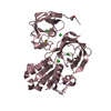
| ||||||||
| Unit cell |
|
-Components
-Protein , 1 types, 3 molecules ABC
| #1: Protein | Mass: 36883.332 Da / Num. of mol.: 3 Source method: isolated from a genetically manipulated source Source: (gene. exp.)  Janthinobacterium sp. (strain J3) (bacteria) Janthinobacterium sp. (strain J3) (bacteria)Strain: J3 / Gene: carAd / Plasmid: pEJ3NAd / Production host: Escherichia coli BL21(DE3) (bacteria) / Strain (production host): BL21(DE3) / References: UniProt: Q84II0 |
|---|
-Non-polymers , 6 types, 196 molecules 


| #2: Chemical | Iron–sulfur cluster Mass: 175.820 Da / Num. of mol.: 3 / Source method: obtained synthetically / Formula: Fe2S2 Mass: 175.820 Da / Num. of mol.: 3 / Source method: obtained synthetically / Formula: Fe2S2#3: Chemical | Iodide Mass: 126.904 Da / Num. of mol.: 3 / Source method: obtained synthetically / Formula: I Mass: 126.904 Da / Num. of mol.: 3 / Source method: obtained synthetically / Formula: I#4: Chemical | ChemComp-CL / Chloride Mass: 35.453 Da / Num. of mol.: 18 / Source method: obtained synthetically / Formula: Cl Mass: 35.453 Da / Num. of mol.: 18 / Source method: obtained synthetically / Formula: Cl#5: Chemical | ChemComp-NI / | Nickel Mass: 58.693 Da / Num. of mol.: 1 / Source method: obtained synthetically / Formula: Ni Mass: 58.693 Da / Num. of mol.: 1 / Source method: obtained synthetically / Formula: Ni#6: Chemical | ChemComp-ACT / | Acetate Mass: 59.044 Da / Num. of mol.: 1 / Source method: obtained synthetically / Formula: C2H3O2 Mass: 59.044 Da / Num. of mol.: 1 / Source method: obtained synthetically / Formula: C2H3O2#7: Water | ChemComp-HOH / | WaterMass: 18.015 Da / Num. of mol.: 170 / Source method: isolated from a natural source / Formula: H2O |
|---|
-Details
| Has ligand of interest | N |
|---|
-Experimental details
-Experiment
| Experiment | Method: X-RAY DIFFRACTION / Number of used crystals: 1 |
|---|
- Sample preparation
| Crystal | Density Matthews: 2.36 Å3/Da / Density % sol: 47.84 % |
|---|---|
| Crystal grow | Temperature: 277 K / Method: vapor diffusion, hanging drop Details: 0.16 mM calcium acetate or magnesium acetate, 8-12%(w/v) PEG 8000, 0.2 M sodium iodide, 0.08 M MES PH range: 6.5-6.7 |
-Data collection
| Diffraction | Mean temperature: 100 K / Serial crystal experiment: N |
|---|---|
| Diffraction source | Source: SYNCHROTRON / Site: Photon Factory / Beamline: AR-NW12A / Wavelength: 1 Å |
| Detector | Type: ADSC QUANTUM 210 / Detector: CCD / Date: May 16, 2006 |
| Radiation | Protocol: SINGLE WAVELENGTH / Monochromatic (M) / Laue (L): M / Scattering type: x-ray |
| Radiation wavelength | Wavelength: 1 Å / Relative weight: 1 |
| Reflection | Resolution: 2.4→44.91 Å / Num. obs: 41796 / % possible obs: 99.7 % / Redundancy: 6.5 % / Rmerge(I) obs: 0.072 / Net I/σ(I): 11.4 |
| Reflection shell | Resolution: 2.4→2.49 Å / Redundancy: 3.9 % / Rmerge(I) obs: 0.412 / Mean I/σ(I) obs: 3 / Num. unique obs: 4118 / % possible all: 97.5 |
- Processing
| Software |
| ||||||||||||||||||||||||||||||||||||||||||||||||||||||||||||||||||||||||||||||||||||||||||||||||||||||||||||||||||||||||||||||||||||||||||||||||||||||||||||||||||||||||||||||||||||||||||||||||||||||||||||||||||||||||||||||||||||||||||||||||||||||||||
|---|---|---|---|---|---|---|---|---|---|---|---|---|---|---|---|---|---|---|---|---|---|---|---|---|---|---|---|---|---|---|---|---|---|---|---|---|---|---|---|---|---|---|---|---|---|---|---|---|---|---|---|---|---|---|---|---|---|---|---|---|---|---|---|---|---|---|---|---|---|---|---|---|---|---|---|---|---|---|---|---|---|---|---|---|---|---|---|---|---|---|---|---|---|---|---|---|---|---|---|---|---|---|---|---|---|---|---|---|---|---|---|---|---|---|---|---|---|---|---|---|---|---|---|---|---|---|---|---|---|---|---|---|---|---|---|---|---|---|---|---|---|---|---|---|---|---|---|---|---|---|---|---|---|---|---|---|---|---|---|---|---|---|---|---|---|---|---|---|---|---|---|---|---|---|---|---|---|---|---|---|---|---|---|---|---|---|---|---|---|---|---|---|---|---|---|---|---|---|---|---|---|---|---|---|---|---|---|---|---|---|---|---|---|---|---|---|---|---|---|---|---|---|---|---|---|---|---|---|---|---|---|---|---|---|---|---|---|---|---|---|---|---|---|---|---|---|---|---|---|---|---|
| Refinement | Method to determine structure: MOLECULAR REPLACEMENT / Resolution: 2.4→43.07 Å / Cor.coef. Fo:Fc: 0.943 / Cor.coef. Fo:Fc free: 0.918 / SU B: 19.452 / SU ML: 0.29 / Cross valid method: THROUGHOUT / σ(F): 0 / ESU R: 0.512 / ESU R Free: 0.302 / Stereochemistry target values: MAXIMUM LIKELIHOOD Details: HYDROGENS HAVE BEEN ADDED IN THE RIDING POSITIONS U VALUES : WITH TLS ADDED
| ||||||||||||||||||||||||||||||||||||||||||||||||||||||||||||||||||||||||||||||||||||||||||||||||||||||||||||||||||||||||||||||||||||||||||||||||||||||||||||||||||||||||||||||||||||||||||||||||||||||||||||||||||||||||||||||||||||||||||||||||||||||||||
| Solvent computation | Ion probe radii: 0.8 Å / Shrinkage radii: 0.8 Å / VDW probe radii: 1.2 Å / Solvent model: MASK | ||||||||||||||||||||||||||||||||||||||||||||||||||||||||||||||||||||||||||||||||||||||||||||||||||||||||||||||||||||||||||||||||||||||||||||||||||||||||||||||||||||||||||||||||||||||||||||||||||||||||||||||||||||||||||||||||||||||||||||||||||||||||||
| Displacement parameters | Biso max: 139.42 Å2 / Biso mean: 70.245 Å2 / Biso min: 32.23 Å2
| ||||||||||||||||||||||||||||||||||||||||||||||||||||||||||||||||||||||||||||||||||||||||||||||||||||||||||||||||||||||||||||||||||||||||||||||||||||||||||||||||||||||||||||||||||||||||||||||||||||||||||||||||||||||||||||||||||||||||||||||||||||||||||
| Refinement step | Cycle: final / Resolution: 2.4→43.07 Å
| ||||||||||||||||||||||||||||||||||||||||||||||||||||||||||||||||||||||||||||||||||||||||||||||||||||||||||||||||||||||||||||||||||||||||||||||||||||||||||||||||||||||||||||||||||||||||||||||||||||||||||||||||||||||||||||||||||||||||||||||||||||||||||
| Refine LS restraints |
| ||||||||||||||||||||||||||||||||||||||||||||||||||||||||||||||||||||||||||||||||||||||||||||||||||||||||||||||||||||||||||||||||||||||||||||||||||||||||||||||||||||||||||||||||||||||||||||||||||||||||||||||||||||||||||||||||||||||||||||||||||||||||||
| LS refinement shell | Resolution: 2.4→2.462 Å / Rfactor Rfree error: 0 / Total num. of bins used: 20
| ||||||||||||||||||||||||||||||||||||||||||||||||||||||||||||||||||||||||||||||||||||||||||||||||||||||||||||||||||||||||||||||||||||||||||||||||||||||||||||||||||||||||||||||||||||||||||||||||||||||||||||||||||||||||||||||||||||||||||||||||||||||||||
| Refinement TLS params. | Method: refined / Refine-ID: X-RAY DIFFRACTION
| ||||||||||||||||||||||||||||||||||||||||||||||||||||||||||||||||||||||||||||||||||||||||||||||||||||||||||||||||||||||||||||||||||||||||||||||||||||||||||||||||||||||||||||||||||||||||||||||||||||||||||||||||||||||||||||||||||||||||||||||||||||||||||
| Refinement TLS group |
|
