[English] 日本語
Yorodumi
- PDB-7c0n: Crystal structure of a self-assembling galactosylated peptide hom... -

+
Open data


ID or keywords:

Loading...

-
Basic information

Entry
Database: PDB / ID: 7c0n
TitleCrystal structure of a self-assembling galactosylated peptide homodimer
ComponentsSelf-assembling galactosylated tyrosine-rich peptide
Keywords
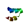
DE NOVO PROTEIN / Self-assembling glycopeptide / tyrosine-rich peptide / O-glycan modification.
Function / homologybeta-D-galactopyranose
Function and homology information
Biological speciessynthetic construct (others)
MethodX-RAY DIFFRACTION / SYNCHROTRON / SAD / Resolution: 1.552 Å
AuthorsHe, C. / Wu, S. / Chi, C. / Zhang, W. / Ma, M. / Lai, L. / Dong, S.
Funding support China, 3items
OrganizationGrant numberCountry
Ministry of Science and Technology (MoST, China)2018YFA0507602 China
Ministry of Science and Technology (MoST, China)2019ZX09301-106 China
National Natural Science Foundation of China (NSFC)21822701 China
CitationJournal: J.Am.Chem.Soc. / Year: 2020
Title: Glycopeptide Self-Assembly Modulated by Glycan Stereochemistry through Glycan-Aromatic Interactions.
Authors: He, C. / Wu, S. / Liu, D. / Chi, C. / Zhang, W. / Ma, M. / Lai, L. / Dong, S.
History
DepositionMay 1, 2020Deposition site: PDBJ / Processing site: PDBJ
Revision 1.0Oct 7, 2020Provider: repository / Type: Initial release
Revision 1.1Oct 21, 2020Group: Database references / Category: citation / citation_author
Item: _citation.journal_volume / _citation.page_first ..._citation.journal_volume / _citation.page_first / _citation.page_last / _citation_author.identifier_ORCID

-
Structure visualization

Structure viewerMolecule:
MolmilJmol/JSmol

Downloads & links

-
Assembly

Deposited unit
A: Self-assembling galactosylated tyrosine-rich peptide
B: Self-assembling galactosylated tyrosine-rich peptide
hetero molecules


Theoretical massNumber of molelcules
Total (without water)2,0045
Polymers1,5482
Non-polymers4563
Water543
1


  • Idetical with deposited unit
  • defined by author&software
  • Evidence: mass spectrometry, The assembly provided by liquid chromatography-mass spectrometry (LC-MS) is monomer.
TypeNameSymmetry operationNumber
identity operation1_555x,y,z1
Buried area960 Å2
ΔGint-16 kcal/mol
Surface area1690 Å2
MethodPISA
Unit cell
Length a, b, c (Å)72.837, 72.837, 72.837
Angle α, β, γ (deg.)90.000, 90.000, 90.000
Int Tables number210
Space group name H-MF4132
Symmetry operation#1: x,y,z
#2: x+1/4,-z+1/4,y+1/4
#3: x+1/4,z+1/4,-y+1/4
#4: z+1/4,y+1/4,-x+1/4
#5: -z+1/4,y+1/4,x+1/4
#6: -y+1/4,x+1/4,z+1/4
#7: y+1/4,-x+1/4,z+1/4
#8: z,x,y
#9: y,z,x
#10: -y,-z,x
#11: z,-x,-y
#12: -y,z,-x
#13: -z,-x,y
#14: -z,x,-y
#15: y,-z,-x
#16: x,-y,-z
#17: -x,y,-z
#18: -x,-y,z
#19: y+1/4,x+1/4,-z+1/4
#20: -y+1/4,-x+1/4,-z+1/4
#21: z+1/4,-y+1/4,x+1/4
#22: -z+1/4,-y+1/4,-x+1/4
#23: -x+1/4,z+1/4,y+1/4
#24: -x+1/4,-z+1/4,-y+1/4
#25: x,y+1/2,z+1/2
#26: x+1/4,-z+3/4,y+3/4
#27: x+1/4,z+3/4,-y+3/4
#28: z+1/4,y+3/4,-x+3/4
#29: -z+1/4,y+3/4,x+3/4
#30: -y+1/4,x+3/4,z+3/4
#31: y+1/4,-x+3/4,z+3/4
#32: z,x+1/2,y+1/2
#33: y,z+1/2,x+1/2
#34: -y,-z+1/2,x+1/2
#35: z,-x+1/2,-y+1/2
#36: -y,z+1/2,-x+1/2
#37: -z,-x+1/2,y+1/2
#38: -z,x+1/2,-y+1/2
#39: y,-z+1/2,-x+1/2
#40: x,-y+1/2,-z+1/2
#41: -x,y+1/2,-z+1/2
#42: -x,-y+1/2,z+1/2
#43: y+1/4,x+3/4,-z+3/4
#44: -y+1/4,-x+3/4,-z+3/4
#45: z+1/4,-y+3/4,x+3/4
#46: -z+1/4,-y+3/4,-x+3/4
#47: -x+1/4,z+3/4,y+3/4
#48: -x+1/4,-z+3/4,-y+3/4
#49: x+1/2,y,z+1/2
#50: x+3/4,-z+1/4,y+3/4
#51: x+3/4,z+1/4,-y+3/4
#52: z+3/4,y+1/4,-x+3/4
#53: -z+3/4,y+1/4,x+3/4
#54: -y+3/4,x+1/4,z+3/4
#55: y+3/4,-x+1/4,z+3/4
#56: z+1/2,x,y+1/2
#57: y+1/2,z,x+1/2
#58: -y+1/2,-z,x+1/2
#59: z+1/2,-x,-y+1/2
#60: -y+1/2,z,-x+1/2
#61: -z+1/2,-x,y+1/2
#62: -z+1/2,x,-y+1/2
#63: y+1/2,-z,-x+1/2
#64: x+1/2,-y,-z+1/2
#65: -x+1/2,y,-z+1/2
#66: -x+1/2,-y,z+1/2
#67: y+3/4,x+1/4,-z+3/4
#68: -y+3/4,-x+1/4,-z+3/4
#69: z+3/4,-y+1/4,x+3/4
#70: -z+3/4,-y+1/4,-x+3/4
#71: -x+3/4,z+1/4,y+3/4
#72: -x+3/4,-z+1/4,-y+3/4
#73: x+1/2,y+1/2,z
#74: x+3/4,-z+3/4,y+1/4
#75: x+3/4,z+3/4,-y+1/4
#76: z+3/4,y+3/4,-x+1/4
#77: -z+3/4,y+3/4,x+1/4
#78: -y+3/4,x+3/4,z+1/4
#79: y+3/4,-x+3/4,z+1/4
#80: z+1/2,x+1/2,y
#81: y+1/2,z+1/2,x
#82: -y+1/2,-z+1/2,x
#83: z+1/2,-x+1/2,-y
#84: -y+1/2,z+1/2,-x
#85: -z+1/2,-x+1/2,y
#86: -z+1/2,x+1/2,-y
#87: y+1/2,-z+1/2,-x
#88: x+1/2,-y+1/2,-z
#89: -x+1/2,y+1/2,-z
#90: -x+1/2,-y+1/2,z
#91: y+3/4,x+3/4,-z+1/4
#92: -y+3/4,-x+3/4,-z+1/4
#93: z+3/4,-y+3/4,x+1/4
#94: -z+3/4,-y+3/4,-x+1/4
#95: -x+3/4,z+3/4,y+1/4
#96: -x+3/4,-z+3/4,-y+1/4

-
Components

#1: Protein/peptide Self-assembling galactosylated tyrosine-rich peptide


Mass: 773.853 Da / Num. of mol.: 2 / Source method: obtained synthetically / Source: (synth.) synthetic construct (others)
#2: Sugar ChemComp-GAL / beta-D-galactopyranose / beta-D-galactose / D-galactose / galactose / Galactose


Type: D-saccharide, beta linking / Mass: 180.156 Da / Num. of mol.: 2 / Source method: isolated from a natural source / Formula: C6H12O6 / Feature type: SUBJECT OF INVESTIGATION
IdentifierTypeProgram
DGalpbCONDENSED IUPAC CARBOHYDRATE SYMBOLGMML 1.0
b-D-galactopyranoseCOMMON NAMEGMML 1.0
b-D-GalpIUPAC CARBOHYDRATE SYMBOLPDB-CARE 1.0
GalSNFG CARBOHYDRATE SYMBOLGMML 1.0
#3: Chemical ChemComp-SO4 / SULFATE ION / Sulfate


Mass: 96.063 Da / Num. of mol.: 1 / Source method: obtained synthetically / Formula: SO4
#4: Water ChemComp-HOH / water / Water


Mass: 18.015 Da / Num. of mol.: 3 / Source method: isolated from a natural source / Formula: H2O
Has ligand of interestY

-
Experimental details

-
Experiment

ExperimentMethod: X-RAY DIFFRACTION / Number of used crystals: 1

-
Sample preparation

CrystalDensity Matthews: 2.6 Å3/Da / Density % sol: 52.71 %
Crystal growTemperature: 293 K / Method: vapor diffusion, sitting drop / pH: 4.2
Details: 40%(w/v) PEG 1000, 0.1 M Sodium phosphate dibasic/Citric acid(pH 4.2), 0.2 M Lithium sulfate

-
Data collection

DiffractionMean temperature: 293 K / Serial crystal experiment: N
Diffraction sourceSource: SYNCHROTRON / Site: SSRF / Beamline: BL17B1 / Wavelength: 1.54191 Å
DetectorType: RAYONIX MX-300 / Detector: CCD / Date: Jun 8, 2019
RadiationProtocol: SINGLE WAVELENGTH / Monochromatic (M) / Laue (L): M / Scattering type: x-ray
Radiation wavelengthWavelength: 1.54191 Å / Relative weight: 1
ReflectionResolution: 1.55→50 Å / Num. obs: 2694 / % possible obs: 100 % / Redundancy: 72.6 % / Rmerge(I) obs: 0.078 / Rsym value: 0.078 / Net I/σ(I): 54.2
Reflection shellResolution: 1.55→1.61 Å / Redundancy: 63 % / Rmerge(I) obs: 0.369 / Mean I/σ(I) obs: 17 / Num. unique obs: 256 / Rsym value: 0.369

-
Processing

Software
NameVersionClassification
PHENIX1.13_2998refinement
HKL-2000data reduction
HKL-2000data scaling
PHENIXphasing
RefinementMethod to determine structure: SAD / Resolution: 1.552→25.752 Å / Cross valid method: THROUGHOUT
RfactorNum. reflection% reflection
Rfree0.2495 118 4.4 %
Rwork0.2344 --
obs-2682 100 %
Displacement parametersBiso max: 52.98 Å2 / Biso min: 10.84 Å2
Refinement stepCycle: final / Resolution: 1.552→25.752 Å
ProteinNucleic acidLigandSolventTotal
Num. atoms107 0 50 3 160
Biso mean--26.31 24.37 -
Num. residues----10
LS refinement shellResolution: 1.552→1.607 Å
RfactorNum. reflection% reflection
Rfree0.2328 --
Rwork0.2326 255 -
obs--100 %

+
About Yorodumi

-
News

-
Feb 9, 2022. New format data for meta-information of EMDB entries

New format data for meta-information of EMDB entries

  • Version 3 of the EMDB header file is now the official format.
  • The previous official version 1.9 will be removed from the archive.

Related info.:EMDB header

External links:wwPDB to switch to version 3 of the EMDB data model

-
Aug 12, 2020. Covid-19 info

Covid-19 info

URL: https://pdbj.org/emnavi/covid19.php

New page: Covid-19 featured information page in EM Navigator.

Related info.:Covid-19 info / Mar 5, 2020. Novel coronavirus structure data

+
Mar 5, 2020. Novel coronavirus structure data

Novel coronavirus structure data

Related info.:Yorodumi Speices / Aug 12, 2020. Covid-19 info

External links:COVID-19 featured content - PDBj / Molecule of the Month (242):Coronavirus Proteases

+
Jan 31, 2019. EMDB accession codes are about to change! (news from PDBe EMDB page)

EMDB accession codes are about to change! (news from PDBe EMDB page)

  • The allocation of 4 digits for EMDB accession codes will soon come to an end. Whilst these codes will remain in use, new EMDB accession codes will include an additional digit and will expand incrementally as the available range of codes is exhausted. The current 4-digit format prefixed with “EMD-” (i.e. EMD-XXXX) will advance to a 5-digit format (i.e. EMD-XXXXX), and so on. It is currently estimated that the 4-digit codes will be depleted around Spring 2019, at which point the 5-digit format will come into force.
  • The EM Navigator/Yorodumi systems omit the EMD- prefix.

Related info.:Q: What is EMD? / ID/Accession-code notation in Yorodumi/EM Navigator

External links:EMDB Accession Codes are Changing Soon! / Contact to PDBj

+
Jul 12, 2017. Major update of PDB

Major update of PDB

  • wwPDB released updated PDB data conforming to the new PDBx/mmCIF dictionary.
  • This is a major update changing the version number from 4 to 5, and with Remediation, in which all the entries are updated.
  • In this update, many items about electron microscopy experimental information are reorganized (e.g. em_software).
  • Now, EM Navigator and Yorodumi are based on the updated data.

External links:wwPDB Remediation / Enriched Model Files Conforming to OneDep Data Standards Now Available in the PDB FTP Archive

-
Yorodumi

Thousand views of thousand structures

  • Yorodumi is a browser for structure data from EMDB, PDB, SASBDB, etc.
  • This page is also the successor to EM Navigator detail page, and also detail information page/front-end page for Omokage search.
  • The word "yorodu" (or yorozu) is an old Japanese word meaning "ten thousand". "mi" (miru) is to see.

Related info.:EMDB / PDB / SASBDB / Comparison of 3 databanks / Yorodumi Search / Aug 31, 2016. New EM Navigator & Yorodumi / Yorodumi Papers / Jmol/JSmol / Function and homology information / Changes in new EM Navigator and Yorodumi

Read more