 Movie
Movie Controller

[English] 日本語
 Yorodumi
- PDB-7c0e: Crystal structure of Azospirillum brasilense L-2-keto-3-deoxyarab... -
+ Open data

- Basic information
| Entry | Database: PDB / ID: 7c0e | ||||||
|---|---|---|---|---|---|---|---|
| Title | Crystal structure of Azospirillum brasilense L-2-keto-3-deoxyarabonate dehydratase (2-oxobutyrate-bound form) | ||||||
 Components Components | L-2-keto-3-deoxyarabonate dehydratase | ||||||
 Keywords Keywords |  LYASE / L-2-keto-3-deoxyarabonate dehydratase LYASE / L-2-keto-3-deoxyarabonate dehydratase | ||||||
| Function / homology |  Function and homology information2-dehydro-3-deoxy-L-arabinonate dehydratase / 2-dehydro-3-deoxy-L-arabinonate dehydratase activity / L-arabinose catabolic process to 2-oxoglutarate / 4-hydroxy-tetrahydrodipicolinate synthase activity / protein homodimerization activity / cytosol Function and homology information2-dehydro-3-deoxy-L-arabinonate dehydratase / 2-dehydro-3-deoxy-L-arabinonate dehydratase activity / L-arabinose catabolic process to 2-oxoglutarate / 4-hydroxy-tetrahydrodipicolinate synthase activity / protein homodimerization activity / cytosolSimilarity search - Function | ||||||
| Biological species |  Azospirillum brasilense (bacteria) Azospirillum brasilense (bacteria) | ||||||
| Method | X-RAY DIFFRACTION / SYNCHROTRON / MOLECULAR REPLACEMENT / Resolution: 2.204 Å | ||||||
 Authors Authors | Watanabe, Y. / Ono, A. / Watanabe, S. | ||||||
 Citation Citation | Journal: Biochemistry / Year: 2020 Title: Biochemical and Structural Characterization of l-2-Keto-3-deoxyarabinonate Dehydratase: A Unique Catalytic Mechanism in the Class I Aldolase Protein Superfamily. Authors: Watanabe, S. / Watanabe, Y. / Nobuchi, R. / Ono, A. | ||||||
| History |
|
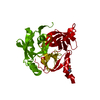
- Structure visualization
| Structure viewer | Molecule: MolmilJmol/JSmol |
|---|
- Downloads & links
-Download
| PDBx/mmCIF format | 7c0e.cif.gz | 692.6 KB | Display | PDBx/mmCIF format |
|---|---|---|---|---|
| PDB format | pdb7c0e.ent.gz | 575.3 KB | Display | PDB format |
| PDBx/mmJSON format | 7c0e.json.gz | Tree view | PDBx/mmJSON format | |
| Others |  Other downloads Other downloads |
-Validation report
| Arichive directory | https://data.pdbj.org/pub/pdb/validation_reports/c0/7c0eftp://data.pdbj.org/pub/pdb/validation_reports/c0/7c0e | HTTPS FTP |
|---|
-Related structure data
| Related structure data |  7c0cSC 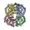 7c0dC S: Starting model for refinement C: citing same article ( |
|---|---|
| Similar structure data |


-Links
 PDBj
PDBj- Assembly
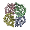
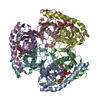
| Deposited unit | 
| ||||||||
|---|---|---|---|---|---|---|---|---|---|
| 1 | 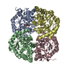
| ||||||||
| 2 | 
| ||||||||
| 3 | 
| ||||||||
| Unit cell |
|
-Components
| #1: Protein | Mass: 35152.902 Da / Num. of mol.: 12 Source method: isolated from a genetically manipulated source Source: (gene. exp.) Azospirillum brasilense (bacteria) / Gene: araD / Production host: Escherichia coli (E. coli)References: UniProt: Q1JUQ0, 2-dehydro-3-deoxy-L-arabinonate dehydratase#2: Water | ChemComp-HOH / | Water Mass: 18.015 Da / Num. of mol.: 1132 / Source method: isolated from a natural source / Formula: H2O Mass: 18.015 Da / Num. of mol.: 1132 / Source method: isolated from a natural source / Formula: H2OHas ligand of interest | Y | |
|---|
-Experimental details
-Experiment
| Experiment | Method: X-RAY DIFFRACTION / Number of used crystals: 1 |
|---|
- Sample preparation
| Crystal | Density % sol: 37.57 % Description: The entry contains friedel pairs in F_plus/minus columns and I_plus/minus columns |
|---|---|
| Crystal grow | Temperature: 293 K / Method: vapor diffusion, sitting drop Details: 0.2 M magnesium chloride, 0.1 M Tris-HCl, 22-27% PEG 3350 PH range: 8.3-8.9 |
-Data collection
| Diffraction | Mean temperature: 100 K / Serial crystal experiment: N | ||||||||||||||||||||||||||||||||||||||||||||||||||||||||||||||||||||||||||||||||||||||||||||||||||||
|---|---|---|---|---|---|---|---|---|---|---|---|---|---|---|---|---|---|---|---|---|---|---|---|---|---|---|---|---|---|---|---|---|---|---|---|---|---|---|---|---|---|---|---|---|---|---|---|---|---|---|---|---|---|---|---|---|---|---|---|---|---|---|---|---|---|---|---|---|---|---|---|---|---|---|---|---|---|---|---|---|---|---|---|---|---|---|---|---|---|---|---|---|---|---|---|---|---|---|---|---|---|
| Diffraction source | Source: SYNCHROTRON / Site: SPring-8  / Beamline: BL45XU / Wavelength: 1 Å / Beamline: BL45XU / Wavelength: 1 Å | ||||||||||||||||||||||||||||||||||||||||||||||||||||||||||||||||||||||||||||||||||||||||||||||||||||
| Detector | Type: DECTRIS PILATUS 6M / Detector: PIXEL / Date: Oct 15, 2019 | ||||||||||||||||||||||||||||||||||||||||||||||||||||||||||||||||||||||||||||||||||||||||||||||||||||
| Radiation | Protocol: SINGLE WAVELENGTH / Monochromatic (M) / Laue (L): M / Scattering type: x-ray | ||||||||||||||||||||||||||||||||||||||||||||||||||||||||||||||||||||||||||||||||||||||||||||||||||||
| Radiation wavelength | Wavelength: 1 Å / Relative weight: 1 | ||||||||||||||||||||||||||||||||||||||||||||||||||||||||||||||||||||||||||||||||||||||||||||||||||||
| Reflection | Resolution: 2.2→47.954 Å / Num. obs: 160387 / % possible obs: 95.9 % / Redundancy: 2.266 % / Biso Wilson estimate: 30.4 Å2 / CC1/2: 0.989 / Rmerge(I) obs: 0.128 / Rrim(I) all: 0.166 / Χ2: 1.146 / Net I/σ(I): 5.54 / Num. measured all: 709648 | ||||||||||||||||||||||||||||||||||||||||||||||||||||||||||||||||||||||||||||||||||||||||||||||||||||
| Reflection shell | Diffraction-ID: 1
|
- Processing
| Software |
| ||||||||||||||||||||||||||||||||||||||||||||||||||||||||||||||||||||||||||||||||||||||||||||||||||||||||||||||||||||||||||||||||||||||||||||||||||||||||||||||||||||||||||||||||||||||||||
|---|---|---|---|---|---|---|---|---|---|---|---|---|---|---|---|---|---|---|---|---|---|---|---|---|---|---|---|---|---|---|---|---|---|---|---|---|---|---|---|---|---|---|---|---|---|---|---|---|---|---|---|---|---|---|---|---|---|---|---|---|---|---|---|---|---|---|---|---|---|---|---|---|---|---|---|---|---|---|---|---|---|---|---|---|---|---|---|---|---|---|---|---|---|---|---|---|---|---|---|---|---|---|---|---|---|---|---|---|---|---|---|---|---|---|---|---|---|---|---|---|---|---|---|---|---|---|---|---|---|---|---|---|---|---|---|---|---|---|---|---|---|---|---|---|---|---|---|---|---|---|---|---|---|---|---|---|---|---|---|---|---|---|---|---|---|---|---|---|---|---|---|---|---|---|---|---|---|---|---|---|---|---|---|---|---|---|---|
| Refinement | Method to determine structure: MOLECULAR REPLACEMENT Starting model: 7C0C Resolution: 2.204→47.954 Å / SU ML: 0.31 / Cross valid method: THROUGHOUT / σ(F): 1.97 / Phase error: 29.34 / Stereochemistry target values: ML Details: The entry contains friedel pairs in F_plus/minus columns and I_plus/minus columns
| ||||||||||||||||||||||||||||||||||||||||||||||||||||||||||||||||||||||||||||||||||||||||||||||||||||||||||||||||||||||||||||||||||||||||||||||||||||||||||||||||||||||||||||||||||||||||||
| Solvent computation | Shrinkage radii: 0.9 Å / VDW probe radii: 1.11 Å / Solvent model: FLAT BULK SOLVENT MODEL | ||||||||||||||||||||||||||||||||||||||||||||||||||||||||||||||||||||||||||||||||||||||||||||||||||||||||||||||||||||||||||||||||||||||||||||||||||||||||||||||||||||||||||||||||||||||||||
| Displacement parameters | Biso max: 87.03 Å2 / Biso mean: 35.5377 Å2 / Biso min: 12.76 Å2 | ||||||||||||||||||||||||||||||||||||||||||||||||||||||||||||||||||||||||||||||||||||||||||||||||||||||||||||||||||||||||||||||||||||||||||||||||||||||||||||||||||||||||||||||||||||||||||
| Refinement step | Cycle: final / Resolution: 2.204→47.954 Å
| ||||||||||||||||||||||||||||||||||||||||||||||||||||||||||||||||||||||||||||||||||||||||||||||||||||||||||||||||||||||||||||||||||||||||||||||||||||||||||||||||||||||||||||||||||||||||||
| Refine LS restraints |
| ||||||||||||||||||||||||||||||||||||||||||||||||||||||||||||||||||||||||||||||||||||||||||||||||||||||||||||||||||||||||||||||||||||||||||||||||||||||||||||||||||||||||||||||||||||||||||
| LS refinement shell | Refine-ID: X-RAY DIFFRACTION / Rfactor Rfree error: 0
|
