 Movie
Movie Controller


+ Open data

- Basic information
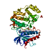
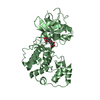
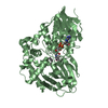
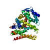
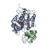
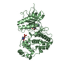
| Entry | Database: PDB / ID: 7byj | ||||||
|---|---|---|---|---|---|---|---|
| Title | Crystal structure of the FERM domain of FRMPD4 | ||||||
 Components Components | FERM and PDZ domain-containing protein 4 | ||||||
 Keywords Keywords |  PROTEIN BINDING / FERM / FRMPD4 / neural scaffold protein PROTEIN BINDING / FERM / FRMPD4 / neural scaffold protein | ||||||
| Function / homology |  Function and homology information Function and homology informationpositive regulation of synapse structural plasticity / phosphatidylinositol-4,5-bisphosphate binding / dendritic spine / cytoskeleton / protein-containing complexSimilarity search - Function | ||||||
| Biological species |  Homo sapiens (human) Homo sapiens (human) | ||||||
| Method | X-RAY DIFFRACTION / SYNCHROTRON / MOLECULAR REPLACEMENT / Resolution: 2.49 Å | ||||||
 Authors Authors | Lin, L. / Wang, M. / Wang, C. / Zhu, J. | ||||||
 Citation Citation | Journal: Biochem.J. / Year: 2020 Title: Structure of the FERM domain of a neural scaffold protein FRMPD4 implicated in X-linked intellectual disability. Authors: Wang, M. / Lin, L. / Shi, Y. / He, L. / Wang, C. / Zhu, J. | ||||||
| History |
|
- Structure visualization
| Structure viewer | Molecule: MolmilJmol/JSmol |
|---|
- Downloads & links
-Download
| PDBx/mmCIF format | 7byj.cif.gz | 80.7 KB | Display | PDBx/mmCIF format |
|---|---|---|---|---|
| PDB format | pdb7byj.ent.gz | 57.2 KB | Display | PDB format |
| PDBx/mmJSON format | 7byj.json.gz | Tree view | PDBx/mmJSON format | |
| Others |  Other downloads Other downloads |
-Validation report
| Arichive directory | https://data.pdbj.org/pub/pdb/validation_reports/by/7byjftp://data.pdbj.org/pub/pdb/validation_reports/by/7byj | HTTPS FTP |
|---|
-Related structure data
| Related structure data |  6e2qS S: Starting model for refinement |
|---|---|
| Similar structure data |


-Links
 PDBj
PDBj

- Assembly
| Deposited unit | 
| ||||||||
|---|---|---|---|---|---|---|---|---|---|
| 1 |
| ||||||||
| Unit cell |
|
-Components
| #1: Protein | Mass: 38562.953 Da / Num. of mol.: 1 Source method: isolated from a genetically manipulated source Source: (gene. exp.) Homo sapiens (human) / Gene: FRMPD4, KIAA0316, PDZD10, PDZK10 / Production host:  Escherichia coli BL21(DE3) (bacteria) / References: UniProt: Q14CM0 Escherichia coli BL21(DE3) (bacteria) / References: UniProt: Q14CM0 |
|---|---|
| #2: Water | ChemComp-HOH / Water Mass: 18.015 Da / Num. of mol.: 10 / Source method: isolated from a natural source / Formula: H2O Mass: 18.015 Da / Num. of mol.: 10 / Source method: isolated from a natural source / Formula: H2O |
-Experimental details
-Experiment
| Experiment | Method: X-RAY DIFFRACTION / Number of used crystals: 1 |
|---|
- Sample preparation
| Crystal | Density Matthews: 2.76 Å3/Da / Density % sol: 55.49 % |
|---|---|
| Crystal grow | Temperature: 289 K / Method: vapor diffusion, sitting drop Details: Lithium citrate tribasic tetrahydrate, Polyethylene glycol 3350 |
-Data collection
| Diffraction | Mean temperature: 100 K / Serial crystal experiment: N | ||||||||||||||||||||||||||||||||||||||||||||||||||||||||||||||||||||||||||||||||||||||||||||||||||||
|---|---|---|---|---|---|---|---|---|---|---|---|---|---|---|---|---|---|---|---|---|---|---|---|---|---|---|---|---|---|---|---|---|---|---|---|---|---|---|---|---|---|---|---|---|---|---|---|---|---|---|---|---|---|---|---|---|---|---|---|---|---|---|---|---|---|---|---|---|---|---|---|---|---|---|---|---|---|---|---|---|---|---|---|---|---|---|---|---|---|---|---|---|---|---|---|---|---|---|---|---|---|
| Diffraction source | Source: SYNCHROTRON / Site: SSRF  / Beamline: BL19U1 / Wavelength: 0.9789 Å / Beamline: BL19U1 / Wavelength: 0.9789 Å | ||||||||||||||||||||||||||||||||||||||||||||||||||||||||||||||||||||||||||||||||||||||||||||||||||||
| Detector | Type: DECTRIS PILATUS 2M / Detector: PIXEL / Date: Oct 10, 2019 | ||||||||||||||||||||||||||||||||||||||||||||||||||||||||||||||||||||||||||||||||||||||||||||||||||||
| Radiation | Protocol: SINGLE WAVELENGTH / Monochromatic (M) / Laue (L): M / Scattering type: x-ray | ||||||||||||||||||||||||||||||||||||||||||||||||||||||||||||||||||||||||||||||||||||||||||||||||||||
| Radiation wavelength | Wavelength: 0.9789 Å / Relative weight: 1 | ||||||||||||||||||||||||||||||||||||||||||||||||||||||||||||||||||||||||||||||||||||||||||||||||||||
| Reflection | Resolution: 2.49→45.3 Å / Num. obs: 16231 / % possible obs: 100 % / Redundancy: 23.56 % / Biso Wilson estimate: 74.598 Å2 / CC1/2: 0.999 / Rmerge(I) obs: 0.087 / Rrim(I) all: 0.089 / Χ2: 0.778 / Net I/σ(I): 23.79 / Num. measured all: 382398 | ||||||||||||||||||||||||||||||||||||||||||||||||||||||||||||||||||||||||||||||||||||||||||||||||||||
| Reflection shell | Diffraction-ID: 1
|
- Processing
| Software |
| ||||||||||||||||||||||||||||||||||||||||||||||||||||||||||||||||||||||||||||||
|---|---|---|---|---|---|---|---|---|---|---|---|---|---|---|---|---|---|---|---|---|---|---|---|---|---|---|---|---|---|---|---|---|---|---|---|---|---|---|---|---|---|---|---|---|---|---|---|---|---|---|---|---|---|---|---|---|---|---|---|---|---|---|---|---|---|---|---|---|---|---|---|---|---|---|---|---|---|---|---|
| Refinement | Method to determine structure: MOLECULAR REPLACEMENT Starting model: 6E2Q Resolution: 2.49→45.3 Å / SU ML: 0.45 / Cross valid method: THROUGHOUT / σ(F): 1.37 / Phase error: 28.76 / Stereochemistry target values: ML
| ||||||||||||||||||||||||||||||||||||||||||||||||||||||||||||||||||||||||||||||
| Solvent computation | Shrinkage radii: 0.9 Å / VDW probe radii: 1.11 Å / Solvent model: FLAT BULK SOLVENT MODEL | ||||||||||||||||||||||||||||||||||||||||||||||||||||||||||||||||||||||||||||||
| Displacement parameters | Biso max: 164.47 Å2 / Biso mean: 73.5663 Å2 / Biso min: 30 Å2 | ||||||||||||||||||||||||||||||||||||||||||||||||||||||||||||||||||||||||||||||
| Refinement step | Cycle: final / Resolution: 2.49→45.3 Å
| ||||||||||||||||||||||||||||||||||||||||||||||||||||||||||||||||||||||||||||||
| LS refinement shell | Refine-ID: X-RAY DIFFRACTION / Rfactor Rfree error: 0 / Total num. of bins used: 12 / % reflection obs: 100 %
|
