 Movie
Movie Controller

[English] 日本語
 Yorodumi
- PDB-7bx9: Purification, characterization and X-ray structure of YhdA-type a... -
+ Open data

- Basic information
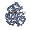
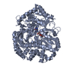
| Entry | Database: PDB / ID: 7bx9 | ||||||
|---|---|---|---|---|---|---|---|
| Title | Purification, characterization and X-ray structure of YhdA-type azoreductase from Bacillus velezensis | ||||||
 Components Components | Azoreductase Azobenzene reductase Azobenzene reductase | ||||||
 Keywords Keywords | OXIDOREDUCTASE / Azo reductase / Azr / FMN / FLAVOPROTEIN | ||||||
| Function / homology | NADPH-dependent FMN reductase-like / NADPH-dependent FMN reductase / Flavoprotein-like superfamily / oxidoreductase activity / FLAVIN MONONUCLEOTIDE / PHOSPHATE ION / Azoreductase Function and homology information Function and homology information | ||||||
| Biological species |  Bacillus amyloliquefaciens (bacteria) Bacillus amyloliquefaciens (bacteria) | ||||||
| Method | X-RAY DIFFRACTION / SYNCHROTRON / MOLECULAR REPLACEMENT / Resolution: 1.38 Å | ||||||
 Authors Authors | Khan, F. / Suguna, K. | ||||||
 Citation Citation | Journal: Proteins / Year: 2021 Title: Purification, characterization, and crystal structure of YhdA-type azoreductase from Bacillus velezensis. Authors: Bafana, A. / Khan, F. / Suguna, K. | ||||||
| History |
|
- Structure visualization
| Structure viewer | Molecule: MolmilJmol/JSmol |
|---|
- Downloads & links
-Download
| PDBx/mmCIF format | 7bx9.cif.gz | 110.8 KB | Display | PDBx/mmCIF format |
|---|---|---|---|---|
| PDB format | pdb7bx9.ent.gz | 69.9 KB | Display | PDB format |
| PDBx/mmJSON format | 7bx9.json.gz | Tree view | PDBx/mmJSON format | |
| Others |  Other downloads Other downloads |
-Validation report
| Arichive directory | https://data.pdbj.org/pub/pdb/validation_reports/bx/7bx9ftp://data.pdbj.org/pub/pdb/validation_reports/bx/7bx9 | HTTPS FTP |
|---|
-Related structure data
| Related structure data |  2gswS S: Starting model for refinement |
|---|---|
| Similar structure data |


-Links
 PDBj
PDBj- Assembly
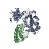
| Deposited unit | 
| ||||||||||||||||||||||||||||||
|---|---|---|---|---|---|---|---|---|---|---|---|---|---|---|---|---|---|---|---|---|---|---|---|---|---|---|---|---|---|---|---|
| 1 | 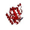
| ||||||||||||||||||||||||||||||
| Unit cell |
| ||||||||||||||||||||||||||||||
| Components on special symmetry positions |
|
-Components
| #1: Protein | Azobenzene reductase Mass: 18632.459 Da / Num. of mol.: 1 Source method: isolated from a genetically manipulated source Source: (gene. exp.) Bacillus amyloliquefaciens (bacteria) / Gene: azr / Production host: Escherichia coli BL21 (bacteria) / Strain (production host): BL21 / References: UniProt: B3VPZ9 | ||||||||
|---|---|---|---|---|---|---|---|---|---|
| #2: Chemical | ChemComp-GOL / Glycerol  Mass: 92.094 Da / Num. of mol.: 5 / Source method: obtained synthetically / Formula: C3H8O3 Mass: 92.094 Da / Num. of mol.: 5 / Source method: obtained synthetically / Formula: C3H8O3#3: Chemical | ChemComp-PO4 / | Phosphate  Mass: 94.971 Da / Num. of mol.: 1 / Source method: obtained synthetically / Formula: PO4 Mass: 94.971 Da / Num. of mol.: 1 / Source method: obtained synthetically / Formula: PO4#4: Chemical | ChemComp-FMN / | Flavin mononucleotide  Mass: 456.344 Da / Num. of mol.: 1 / Source method: isolated from a natural source / Formula: C17H21N4O9P / Feature type: SUBJECT OF INVESTIGATION Mass: 456.344 Da / Num. of mol.: 1 / Source method: isolated from a natural source / Formula: C17H21N4O9P / Feature type: SUBJECT OF INVESTIGATION#5: Water | ChemComp-HOH / | Water Mass: 18.015 Da / Num. of mol.: 220 / Source method: isolated from a natural source / Formula: H2O Mass: 18.015 Da / Num. of mol.: 220 / Source method: isolated from a natural source / Formula: H2OHas ligand of interest | Y | |
-Experimental details
-Experiment
| Experiment | Method: X-RAY DIFFRACTION / Number of used crystals: 1 |
|---|
- Sample preparation
| Crystal | Density Matthews: 3.02 Å3/Da / Density % sol: 59.3 % |
|---|---|
| Crystal grow | Temperature: 289 K / Method: batch mode Details: 0.05 M Potassium phosphate monobasic, 20% w/v Polyethylene glycol 8,000 |
-Data collection
| Diffraction | Mean temperature: 100 K / Serial crystal experiment: N |
|---|---|
| Diffraction source | Source: SYNCHROTRON / Site: ESRF  / Beamline: BM14 / Wavelength: 0.9537 Å / Beamline: BM14 / Wavelength: 0.9537 Å |
| Detector | Type: MARMOSAIC 225 mm CCD / Detector: CCD / Date: Apr 4, 2015 |
| Radiation | Protocol: SINGLE WAVELENGTH / Monochromatic (M) / Laue (L): M / Scattering type: x-ray |
| Radiation wavelength | Wavelength: 0.9537 Å / Relative weight: 1 |
| Reflection | Resolution: 1.38→36.71 Å / Num. obs: 51008 / % possible obs: 100 % / Redundancy: 5.4 % / Biso Wilson estimate: 11.69 Å2 / CC1/2: 0.996 / Net I/σ(I): 10 |
| Reflection shell | Resolution: 1.38→1.41 Å / Mean I/σ(I) obs: 2 / Num. unique obs: 2504 / CC1/2: 0.792 |
- Processing
| Software |
| ||||||||||||||||||||||||||||||||||||||||||||||||||||||||||||||||||||||||||||||||||||||||||||||||||||||||||||||||||||||||||||||||||||||||||||
|---|---|---|---|---|---|---|---|---|---|---|---|---|---|---|---|---|---|---|---|---|---|---|---|---|---|---|---|---|---|---|---|---|---|---|---|---|---|---|---|---|---|---|---|---|---|---|---|---|---|---|---|---|---|---|---|---|---|---|---|---|---|---|---|---|---|---|---|---|---|---|---|---|---|---|---|---|---|---|---|---|---|---|---|---|---|---|---|---|---|---|---|---|---|---|---|---|---|---|---|---|---|---|---|---|---|---|---|---|---|---|---|---|---|---|---|---|---|---|---|---|---|---|---|---|---|---|---|---|---|---|---|---|---|---|---|---|---|---|---|---|---|
| Refinement | Method to determine structure: MOLECULAR REPLACEMENT Starting model: 2GSW Resolution: 1.38→30.97 Å / SU ML: 0.1158 / Cross valid method: FREE R-VALUE / σ(F): 1.35 / Phase error: 12.2304 Stereochemistry target values: GeoStd + Monomer Library + CDL v1.2
| ||||||||||||||||||||||||||||||||||||||||||||||||||||||||||||||||||||||||||||||||||||||||||||||||||||||||||||||||||||||||||||||||||||||||||||
| Solvent computation | Shrinkage radii: 0.9 Å / VDW probe radii: 1.11 Å / Solvent model: FLAT BULK SOLVENT MODEL | ||||||||||||||||||||||||||||||||||||||||||||||||||||||||||||||||||||||||||||||||||||||||||||||||||||||||||||||||||||||||||||||||||||||||||||
| Displacement parameters | Biso mean: 18.53 Å2 | ||||||||||||||||||||||||||||||||||||||||||||||||||||||||||||||||||||||||||||||||||||||||||||||||||||||||||||||||||||||||||||||||||||||||||||
| Refinement step | Cycle: LAST / Resolution: 1.38→30.97 Å
| ||||||||||||||||||||||||||||||||||||||||||||||||||||||||||||||||||||||||||||||||||||||||||||||||||||||||||||||||||||||||||||||||||||||||||||
| Refine LS restraints |
| ||||||||||||||||||||||||||||||||||||||||||||||||||||||||||||||||||||||||||||||||||||||||||||||||||||||||||||||||||||||||||||||||||||||||||||
| LS refinement shell |
|
