 Movie
Movie Controller


+ Open data

- Basic information
| Entry | Database: PDB / ID: 7bvw | ||||||
|---|---|---|---|---|---|---|---|
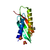
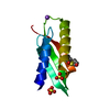
| Title | Crystal structure of the RING-H2 domain of Arabidopsis RMR1 | ||||||
 Components Components | AT5G66160 protein | ||||||
 Keywords Keywords |  TRANSPORT PROTEIN / C3H2C3 / ring finger / E3 ligase TRANSPORT PROTEIN / C3H2C3 / ring finger / E3 ligase | ||||||
| Function / homology | Ring finger domain / Ring finger / Zinc finger RING-type profile. / Zinc finger, RING-type / membrane => GO:0016020 / Zinc finger, RING/FYVE/PHD-type / AT5G66160 protein Function and homology information Function and homology information | ||||||
| Biological species |  Arabidopsis thaliana (thale cress) Arabidopsis thaliana (thale cress) | ||||||
| Method | X-RAY DIFFRACTION / MOLECULAR REPLACEMENT / Resolution: 2.1 Å | ||||||
 Authors Authors | Chen, S. / Wong, K.B. | ||||||
 Citation Citation | Journal: To Be Published Title: The RING-finger of AtRMR1 (Arabidopsis receptor-homology-transmembrane-RING-H2 sorting receptor 1) is an E3 ligase that mediate its trafficking Authors: Chen, S. / Zeng, Y.L. / Wong, H.Y. / Yang, L. / Luo, F. / Jiang, L.W. / Wong, K.B. | ||||||
| History |
|
- Structure visualization
| Structure viewer | Molecule: MolmilJmol/JSmol |
|---|
- Downloads & links
-Download
| PDBx/mmCIF format | 7bvw.cif.gz | 79.6 KB | Display | PDBx/mmCIF format |
|---|---|---|---|---|
| PDB format | pdb7bvw.ent.gz | 57.6 KB | Display | PDB format |
| PDBx/mmJSON format | 7bvw.json.gz | Tree view | PDBx/mmJSON format | |
| Others |  Other downloads Other downloads |
-Validation report
| Arichive directory | https://data.pdbj.org/pub/pdb/validation_reports/bv/7bvwftp://data.pdbj.org/pub/pdb/validation_reports/bv/7bvw | HTTPS FTP |
|---|
-Related structure data
| Related structure data |  4v3kS S: Starting model for refinement |
|---|---|
| Similar structure data |


-Links
 PDBj
PDBj

- Assembly
| Deposited unit | 
| ||||||||
|---|---|---|---|---|---|---|---|---|---|
| 1 | 
| ||||||||
| 2 | 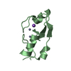
| ||||||||
| Unit cell |
| ||||||||
| Components on special symmetry positions |
|
-Components
| #1: Protein | Mass: 9220.559 Da / Num. of mol.: 2 Source method: isolated from a genetically manipulated source Source: (gene. exp.) Arabidopsis thaliana (thale cress) / Gene: At5g66160 / Production host:  Escherichia coli (E. coli) / Strain (production host): SoluBL21 / References: UniProt: C0Z2X4 Escherichia coli (E. coli) / Strain (production host): SoluBL21 / References: UniProt: C0Z2X4#2: Chemical | ChemComp-ZN /   Mass: 65.409 Da / Num. of mol.: 5 / Source method: obtained synthetically / Formula: Zn / Feature type: SUBJECT OF INVESTIGATION Mass: 65.409 Da / Num. of mol.: 5 / Source method: obtained synthetically / Formula: Zn / Feature type: SUBJECT OF INVESTIGATION#3: Chemical | ChemComp-NA / |   Mass: 22.990 Da / Num. of mol.: 1 / Source method: obtained synthetically / Formula: Na Mass: 22.990 Da / Num. of mol.: 1 / Source method: obtained synthetically / Formula: Na#4: Water | ChemComp-HOH / | Water Mass: 18.015 Da / Num. of mol.: 73 / Source method: isolated from a natural source / Formula: H2O Mass: 18.015 Da / Num. of mol.: 73 / Source method: isolated from a natural source / Formula: H2OHas ligand of interest | Y | |
|---|
-Experimental details
-Experiment
| Experiment | Method: X-RAY DIFFRACTION / Number of used crystals: 1 |
|---|
- Sample preparation
| Crystal | Density Matthews: 1.8 Å3/Da / Density % sol: 31.67 % |
|---|---|
| Crystal grow | Temperature: 289 K / Method: vapor diffusion, hanging drop / pH: 5.5 Details: 0.2 M Ammonium acetate, 0.1 M BIS-TRIS pH 5.5, 20% w/v Polyethylene glycol 3,350 |
-Data collection
| Diffraction | Mean temperature: 93 K / Serial crystal experiment: N |
|---|---|
| Diffraction source | Source: ROTATING ANODE / Type: RIGAKU FR-E+ SUPERBRIGHT / Wavelength: 1.54 Å |
| Detector | Type: RIGAKU RAXIS IV++ / Detector: IMAGE PLATE / Date: Jan 4, 2018 |
| Radiation | Protocol: SINGLE WAVELENGTH / Monochromatic (M) / Laue (L): M / Scattering type: x-ray |
| Radiation wavelength | Wavelength: 1.54 Å / Relative weight: 1 |
| Reflection | Resolution: 2.1→37.72 Å / Num. obs: 7849 / % possible obs: 98.5 % / Redundancy: 3.6 % / CC1/2: 0.992 / Rmerge(I) obs: 0.098 / Rpim(I) all: 0.061 / Rrim(I) all: 0.116 / Net I/σ(I): 9 / Num. measured all: 27884 |
| Reflection shell | Resolution: 2.1→2.16 Å / Redundancy: 3.4 % / Rmerge(I) obs: 0.213 / Num. unique obs: 650 / CC1/2: 0.95 / Rpim(I) all: 0.135 / Rrim(I) all: 0.253 / % possible all: 97.4 |
- Processing
| Software |
| ||||||||||||||||||||||||||||||||||||||||
|---|---|---|---|---|---|---|---|---|---|---|---|---|---|---|---|---|---|---|---|---|---|---|---|---|---|---|---|---|---|---|---|---|---|---|---|---|---|---|---|---|---|
| Refinement | Method to determine structure: MOLECULAR REPLACEMENT Starting model: 4V3K Resolution: 2.1→32.23 Å / SU ML: 0.22 / Cross valid method: THROUGHOUT / σ(F): 1.36 / Phase error: 25.31
| ||||||||||||||||||||||||||||||||||||||||
| Solvent computation | Shrinkage radii: 0.9 Å / VDW probe radii: 1.11 Å | ||||||||||||||||||||||||||||||||||||||||
| Displacement parameters | Biso max: 87.05 Å2 / Biso mean: 27.392 Å2 / Biso min: 12.33 Å2 | ||||||||||||||||||||||||||||||||||||||||
| Refinement step | Cycle: final / Resolution: 2.1→32.23 Å
| ||||||||||||||||||||||||||||||||||||||||
| LS refinement shell | Refine-ID: X-RAY DIFFRACTION / Rfactor Rfree error: 0
| ||||||||||||||||||||||||||||||||||||||||
| Refinement TLS params. | Method: refined / Origin x: 1.4047 Å / Origin y: 5.8871 Å / Origin z: 94.4639 Å
| ||||||||||||||||||||||||||||||||||||||||
| Refinement TLS group |
|
