 Movie
Movie Controller


+ Open data

- Basic information
| Entry | Database: PDB / ID: 7bq9 | ||||||||||||
|---|---|---|---|---|---|---|---|---|---|---|---|---|---|
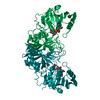
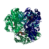
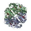
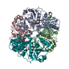
| Title | Crystal structure of ASFV p15 | ||||||||||||
 Components Components | 60 kDa polyprotein | ||||||||||||
 Keywords Keywords |  STRUCTURAL PROTEIN / ASFV / Core-shell / p15 STRUCTURAL PROTEIN / ASFV / Core-shell / p15 | ||||||||||||
| Function / homology | viral capsid assembly / virion component / host cell perinuclear region of cytoplasm / 60 kDa polyprotein / Polyprotein pp62 Function and homology information Function and homology information | ||||||||||||
| Biological species |   African swine fever virus African swine fever virus | ||||||||||||
| Method | X-RAY DIFFRACTION / SYNCHROTRON / SAD / Resolution: 2.612 Å | ||||||||||||
 Authors Authors | Fu, D. / Chen, C. / Guo, Y. | ||||||||||||
| Funding support |  China, 3items China, 3items
| ||||||||||||
 Citation Citation | Journal: Protein Cell / Year: 2020 Title: Structure of a bifunctional membrane-DNA binding protein, African swine fever virus p15 Authors: Fu, D. / Guo, Y. | ||||||||||||
| History |
|
- Structure visualization
| Structure viewer | Molecule: MolmilJmol/JSmol |
|---|
- Downloads & links
-Download
| PDBx/mmCIF format | 7bq9.cif.gz | 133.3 KB | Display | PDBx/mmCIF format |
|---|---|---|---|---|
| PDB format | pdb7bq9.ent.gz | 109.6 KB | Display | PDB format |
| PDBx/mmJSON format | 7bq9.json.gz | Tree view | PDBx/mmJSON format | |
| Others |  Other downloads Other downloads |
-Validation report
| Arichive directory | https://data.pdbj.org/pub/pdb/validation_reports/bq/7bq9ftp://data.pdbj.org/pub/pdb/validation_reports/bq/7bq9 | HTTPS FTP |
|---|
-Related structure data
| Similar structure data |
|---|


-Links
 PDBj
PDBj- Assembly
| Deposited unit | 
| ||||||||
|---|---|---|---|---|---|---|---|---|---|
| 1 | 
| ||||||||
| Unit cell |
|
-Components
| #1: Protein | Mass: 21807.086 Da / Num. of mol.: 2 Source method: isolated from a genetically manipulated source Details: SF file contains Friedel pairs. / Source: (gene. exp.) African swine fever virus / Gene: CP530R / Production host:  Drosophila melanogaster (fruit fly) / References: UniProt: A0A0A1DY09, UniProt: Q65179*PLUS Drosophila melanogaster (fruit fly) / References: UniProt: A0A0A1DY09, UniProt: Q65179*PLUS |
|---|
-Experimental details
-Experiment
| Experiment | Method: X-RAY DIFFRACTION / Number of used crystals: 1 |
|---|
- Sample preparation
| Crystal | Density Matthews: 3.04 Å3/Da / Density % sol: 59.53 % |
|---|---|
| Crystal grow | Temperature: 289 K / Method: vapor diffusion, hanging drop Details: 2.0 M Ammonium sulfate, 0.08M BIS-TRIS propane PH 7.0, 0.22M Sodium malonate PH 7.0, 0.02M HEPES PH7.0, 0.1% v/v Jeffamine ED-2001 PH 7.0 |
-Data collection
| Diffraction | Mean temperature: 100 K / Serial crystal experiment: N |
|---|---|
| Diffraction source | Source: SYNCHROTRON / Site: SSRF / Beamline: BL17U1 / Wavelength: 1.07146 Å |
| Detector | Type: DECTRIS EIGER X 16M / Detector: PIXEL / Date: Jun 14, 2019 |
| Radiation | Protocol: SINGLE WAVELENGTH / Monochromatic (M) / Laue (L): M / Scattering type: x-ray |
| Radiation wavelength | Wavelength: 1.07146 Å / Relative weight: 1 |
| Reflection | Resolution: 2.6→50 Å / Num. obs: 31107 / % possible obs: 99.6 % / Redundancy: 40.2 % / CC1/2: 1 / Net I/σ(I): 30.73 |
| Reflection shell | Resolution: 2.61→2.76 Å / Num. unique obs: 4975 / CC1/2: 0.511 |
- Processing
| Software |
| ||||||||||||||||||||||||||||||||||||||||||||||||||||||||||||||||||||||||||||||||||||||||||||||||||||||||||||||||||||||||||||||||||||||||||
|---|---|---|---|---|---|---|---|---|---|---|---|---|---|---|---|---|---|---|---|---|---|---|---|---|---|---|---|---|---|---|---|---|---|---|---|---|---|---|---|---|---|---|---|---|---|---|---|---|---|---|---|---|---|---|---|---|---|---|---|---|---|---|---|---|---|---|---|---|---|---|---|---|---|---|---|---|---|---|---|---|---|---|---|---|---|---|---|---|---|---|---|---|---|---|---|---|---|---|---|---|---|---|---|---|---|---|---|---|---|---|---|---|---|---|---|---|---|---|---|---|---|---|---|---|---|---|---|---|---|---|---|---|---|---|---|---|---|---|---|
| Refinement | Method to determine structure: SAD / Resolution: 2.612→47.658 Å / SU ML: 0.41 / Cross valid method: THROUGHOUT / σ(F): 1.35 / Phase error: 24.46
| ||||||||||||||||||||||||||||||||||||||||||||||||||||||||||||||||||||||||||||||||||||||||||||||||||||||||||||||||||||||||||||||||||||||||||
| Solvent computation | Shrinkage radii: 0.9 Å / VDW probe radii: 1.11 Å | ||||||||||||||||||||||||||||||||||||||||||||||||||||||||||||||||||||||||||||||||||||||||||||||||||||||||||||||||||||||||||||||||||||||||||
| Displacement parameters | Biso max: 175.73 Å2 / Biso mean: 94.1074 Å2 / Biso min: 52.14 Å2 | ||||||||||||||||||||||||||||||||||||||||||||||||||||||||||||||||||||||||||||||||||||||||||||||||||||||||||||||||||||||||||||||||||||||||||
| Refinement step | Cycle: final / Resolution: 2.612→47.658 Å
| ||||||||||||||||||||||||||||||||||||||||||||||||||||||||||||||||||||||||||||||||||||||||||||||||||||||||||||||||||||||||||||||||||||||||||
| LS refinement shell | Refine-ID: X-RAY DIFFRACTION / Rfactor Rfree error: 0
|
