 Movie
Movie Controller

[English] 日本語
 Yorodumi
- PDB-7bpc: Crystal structure of 2, 3-dihydroxybenzoic acid decarboxylase fro... -
+ Open data

- Basic information
| Entry | Database: PDB / ID: 7bpc | ||||||
|---|---|---|---|---|---|---|---|
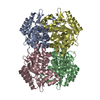
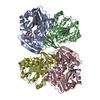
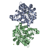
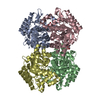
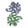
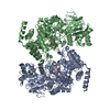
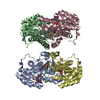
| Title | Crystal structure of 2, 3-dihydroxybenzoic acid decarboxylase from Fusarium oxysporum in complex with 2,5-DHBA | ||||||
 Components Components | 2,3-dihydroxybenzoate decarboxylase | ||||||
 Keywords Keywords |  BIOSYNTHETIC PROTEIN / 2 / 3-dihydroxybenzoic acid decarboxylase BIOSYNTHETIC PROTEIN / 2 / 3-dihydroxybenzoic acid decarboxylase | ||||||
| Function / homology |  Function and homology information Function and homology informationorganic substance metabolic process / carboxy-lyase activity / hydrolase activitySimilarity search - Function | ||||||
| Biological species |   Fusarium oxysporum (fungus) Fusarium oxysporum (fungus) | ||||||
| Method | X-RAY DIFFRACTION / SYNCHROTRON / MOLECULAR REPLACEMENT / Resolution: 2.45 Å | ||||||
 Authors Authors | Song, M.K. / Feng, J.H. / Liu, W.D. / Wu, Q.Q. / Zhu, D.M. | ||||||
 Citation Citation | Journal: Chembiochem / Year: 2020 Title: 2,3-Dihydroxybenzoic Acid Decarboxylase from Fusarium oxysporum: Crystal Structures and Substrate Recognition Mechanism. Authors: Song, M. / Zhang, X. / Liu, W. / Feng, J. / Cui, Y. / Yao, P. / Wang, M. / Guo, R.T. / Wu, Q. / Zhu, D. | ||||||
| History |
|
- Structure visualization
| Structure viewer | Molecule: MolmilJmol/JSmol |
|---|
- Downloads & links
-Download
| PDBx/mmCIF format | 7bpc.cif.gz | 276.3 KB | Display | PDBx/mmCIF format |
|---|---|---|---|---|
| PDB format | pdb7bpc.ent.gz | 221.5 KB | Display | PDB format |
| PDBx/mmJSON format | 7bpc.json.gz | Tree view | PDBx/mmJSON format | |
| Others |  Other downloads Other downloads |
-Validation report
| Arichive directory | https://data.pdbj.org/pub/pdb/validation_reports/bp/7bpcftp://data.pdbj.org/pub/pdb/validation_reports/bp/7bpc | HTTPS FTP |
|---|
-Related structure data
| Related structure data |  6m53C  7bp1C  2dvtS S: Starting model for refinement C: citing same article ( |
|---|---|
| Similar structure data |
-Links
 PDBj
PDBj

- Assembly
| Deposited unit | 
| ||||||||||||||||||||||||||||||||||||||||||||||||||||||||||||||||||||||||||||||||||||||||
|---|---|---|---|---|---|---|---|---|---|---|---|---|---|---|---|---|---|---|---|---|---|---|---|---|---|---|---|---|---|---|---|---|---|---|---|---|---|---|---|---|---|---|---|---|---|---|---|---|---|---|---|---|---|---|---|---|---|---|---|---|---|---|---|---|---|---|---|---|---|---|---|---|---|---|---|---|---|---|---|---|---|---|---|---|---|---|---|---|---|
| 1 |
| ||||||||||||||||||||||||||||||||||||||||||||||||||||||||||||||||||||||||||||||||||||||||
| Unit cell |
| ||||||||||||||||||||||||||||||||||||||||||||||||||||||||||||||||||||||||||||||||||||||||
| Noncrystallographic symmetry (NCS) | NCS domain:
NCS domain segments: Ens-ID: 1
|