 Movie
Movie Controller


+ Open data

- Basic information
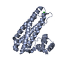
| Entry | Database: PDB / ID: 7bha | ||||||
|---|---|---|---|---|---|---|---|
| Title | Escherichia coli YtfE | ||||||
 Components Components | Iron-sulfur cluster repair protein YtfE | ||||||
 Keywords Keywords | METAL BINDING PROTEIN / di-iron | ||||||
| Function / homology |  Function and homology information Function and homology informationprotein repair / response to nitrosative stress / response to oxidative stress / iron ion binding /  cytosol cytosolSimilarity search - Function | ||||||
| Biological species |  Escherichia coli (E. coli) Escherichia coli (E. coli) | ||||||
| Method | X-RAY DIFFRACTION / SYNCHROTRON / MOLECULAR REPLACEMENT / Resolution: 2.19 Å | ||||||
 Authors Authors | Silva, L.S.O. / Matias, P.M. / Romao, C.V. / Saraiva, L.M. | ||||||
| Funding support |  Portugal, 1items Portugal, 1items
| ||||||
 Citation Citation | Journal: Front Microbiol / Year: 2021 Title: Structural Basis of RICs Iron Donation for Iron-Sulfur Cluster Biogenesis. Authors: Silva, L.S.O. / Matias, P.M. / Romao, C.V. / Saraiva, L.M. | ||||||
| History |
|
- Structure visualization
| Structure viewer | Molecule: MolmilJmol/JSmol |
|---|
- Downloads & links
-Download
| PDBx/mmCIF format | 7bha.cif.gz | 211.8 KB | Display | PDBx/mmCIF format |
|---|---|---|---|---|
| PDB format | pdb7bha.ent.gz | 139.3 KB | Display | PDB format |
| PDBx/mmJSON format | 7bha.json.gz | Tree view | PDBx/mmJSON format | |
| Others |  Other downloads Other downloads |
-Validation report
| Arichive directory | https://data.pdbj.org/pub/pdb/validation_reports/bh/7bhaftp://data.pdbj.org/pub/pdb/validation_reports/bh/7bha | HTTPS FTP |
|---|
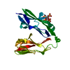
-Related structure data
| Related structure data |  7bhbC  7bhcC 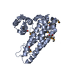 5fnnS S: Starting model for refinement C: citing same article ( |
|---|---|
| Similar structure data |


-Links
 PDBj
PDBj

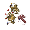
- Assembly
| Deposited unit | 
| ||||||||||||
|---|---|---|---|---|---|---|---|---|---|---|---|---|---|
| 1 | 
| ||||||||||||
| 2 | 
| ||||||||||||
| Unit cell |
| ||||||||||||
| Noncrystallographic symmetry (NCS) | NCS domain:
NCS oper: (Code: givenMatrix: (0.937154934584, -0.0127426509258, 0.348680732808), (-0.0132090181169, -0.999912216244, -0.00104002253238), (0.348663376948, -0.00363106786838, -0.937240985511)Vector: - ...NCS oper: (Code: given Matrix: (0.937154934584, -0.0127426509258, 0.348680732808), Vector : |
-Components
| #1: Protein | Mass: 24720.316 Da / Num. of mol.: 2 / Mutation: C30A C31A Source method: isolated from a genetically manipulated source Source: (gene. exp.) Escherichia coli (strain K12) (bacteria)Strain: K12 / Gene: ytfE, b4209, JW4167 Production host: Escherichia coli 'BL21-Gold(DE3)pLysS AG' (bacteria)References: UniProt: P69506 #2: Chemical | ChemComp-FE / Iron  Mass: 55.845 Da / Num. of mol.: 4 / Source method: obtained synthetically / Formula: Fe Mass: 55.845 Da / Num. of mol.: 4 / Source method: obtained synthetically / Formula: Fe#3: Chemical | Oxygen  Mass: 15.999 Da / Num. of mol.: 2 / Source method: obtained synthetically / Formula: O Mass: 15.999 Da / Num. of mol.: 2 / Source method: obtained synthetically / Formula: O#4: Chemical | ChemComp-GOL / | Glycerol  Mass: 92.094 Da / Num. of mol.: 1 / Source method: obtained synthetically / Formula: C3H8O3 Mass: 92.094 Da / Num. of mol.: 1 / Source method: obtained synthetically / Formula: C3H8O3#5: Water | ChemComp-HOH / | Water Mass: 18.015 Da / Num. of mol.: 83 / Source method: isolated from a natural source / Formula: H2O Mass: 18.015 Da / Num. of mol.: 83 / Source method: isolated from a natural source / Formula: H2OHas ligand of interest | N | |
|---|
-Experimental details
-Experiment
| Experiment | Method: X-RAY DIFFRACTION / Number of used crystals: 1 |
|---|
- Sample preparation
| Crystal | Density Matthews: 2.19 Å3/Da / Density % sol: 54.5 % / Description: needle shapped |
|---|---|
| Crystal grow | Temperature: 298.15 K / Method: vapor diffusion, hanging drop / pH: 7.5 / Details: Tris-HCl, PEG 4000, magnesium chloride |
-Data collection
| Diffraction | Mean temperature: 80 K / Serial crystal experiment: N |
|---|---|
| Diffraction source | Source: SYNCHROTRON / Site: ALBA  / Beamline: XALOC / Wavelength: 1.7314 Å / Beamline: XALOC / Wavelength: 1.7314 Å |
| Detector | Type: DECTRIS PILATUS 6M / Detector: PIXEL / Date: Mar 31, 2019 |
| Radiation | Protocol: SINGLE WAVELENGTH / Monochromatic (M) / Laue (L): M / Scattering type: x-ray |
| Radiation wavelength | Wavelength: 1.7314 Å / Relative weight: 1 |
| Reflection | Resolution: 2.188→59.05 Å / Num. obs: 23568 / % possible obs: 87 % / Redundancy: 6.4 % / Biso Wilson estimate: 37.78 Å2 / CC1/2: 0.985 / Rmerge(I) obs: 0.147 / Rpim(I) all: 0.062 / Rrim(I) all: 0.16 / Net I/σ(I): 9.8 |
| Reflection shell | Resolution: 2.188→2.188 Å / Rmerge(I) obs: 1.548 / Num. unique obs: 549 / CC1/2: 0.354 / % possible all: 40.2 |
- Processing
| Software |
| |||||||||||||||||||||||||||||||||||||||||||||||||||||||||||||||||||||||||||||||||||||||||||||||||||||||||||||||||||||||
|---|---|---|---|---|---|---|---|---|---|---|---|---|---|---|---|---|---|---|---|---|---|---|---|---|---|---|---|---|---|---|---|---|---|---|---|---|---|---|---|---|---|---|---|---|---|---|---|---|---|---|---|---|---|---|---|---|---|---|---|---|---|---|---|---|---|---|---|---|---|---|---|---|---|---|---|---|---|---|---|---|---|---|---|---|---|---|---|---|---|---|---|---|---|---|---|---|---|---|---|---|---|---|---|---|---|---|---|---|---|---|---|---|---|---|---|---|---|---|---|---|
| Refinement | Method to determine structure: MOLECULAR REPLACEMENT Starting model: 5FNN Resolution: 2.19→59.05 Å / SU ML: 0.2773 / Cross valid method: FREE R-VALUE / σ(F): 1.34 / Phase error: 30.3811 Stereochemistry target values: GeoStd + Monomer Library + CDL v1.2 Details: Structure refined with anomalous data
| |||||||||||||||||||||||||||||||||||||||||||||||||||||||||||||||||||||||||||||||||||||||||||||||||||||||||||||||||||||||
| Solvent computation | Shrinkage radii: 0.9 Å / VDW probe radii: 1.11 Å / Solvent model: FLAT BULK SOLVENT MODEL | |||||||||||||||||||||||||||||||||||||||||||||||||||||||||||||||||||||||||||||||||||||||||||||||||||||||||||||||||||||||
| Displacement parameters | Biso mean: 57.67 Å2 | |||||||||||||||||||||||||||||||||||||||||||||||||||||||||||||||||||||||||||||||||||||||||||||||||||||||||||||||||||||||
| Refinement step | Cycle: LAST / Resolution: 2.19→59.05 Å
| |||||||||||||||||||||||||||||||||||||||||||||||||||||||||||||||||||||||||||||||||||||||||||||||||||||||||||||||||||||||
| Refine LS restraints |
| |||||||||||||||||||||||||||||||||||||||||||||||||||||||||||||||||||||||||||||||||||||||||||||||||||||||||||||||||||||||
| LS refinement shell |
|
