 Movie
Movie Controller


+ Open data

- Basic information
| Entry | Database: PDB / ID: 7bh8 | ||||||||||||
|---|---|---|---|---|---|---|---|---|---|---|---|---|---|
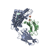
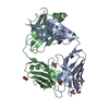
| Title | 3H4-Fab HLA-E-VL9 co-complex | ||||||||||||
 Components Components |
| ||||||||||||
 Keywords Keywords |  IMMUNE SYSTEM / Antibody Fab Fragment MHC class I molecule Human leukocyte antigen E IMMUNE SYSTEM / Antibody Fab Fragment MHC class I molecule Human leukocyte antigen E | ||||||||||||
| Function / homology |  Function and homology information Function and homology informationpositive regulation of antibody-dependent cellular cytotoxicity / regulation of natural killer cell mediated immunity / positive regulation of TRAIL production / antigen processing and presentation of exogenous peptide antigen via MHC class Ib / MHC class Ib protein complex / positive regulation of natural killer cell mediated immunity / positive regulation of natural killer cell cytokine production / natural killer cell tolerance induction / natural killer cell lectin-like receptor binding / negative regulation of natural killer cell activation ...positive regulation of antibody-dependent cellular cytotoxicity / regulation of natural killer cell mediated immunity / positive regulation of TRAIL production / antigen processing and presentation of exogenous peptide antigen via MHC class Ib / MHC class Ib protein complex / positive regulation of natural killer cell mediated immunity / positive regulation of natural killer cell cytokine production / natural killer cell tolerance induction / natural killer cell lectin-like receptor binding / negative regulation of natural killer cell activation / positive regulation of natural killer cell activation / negative regulation of natural killer cell mediated cytotoxicity / positive regulation of natural killer cell mediated cytotoxicity / positive regulation of interleukin-13 production / positive regulation of natural killer cell proliferation / positive regulation of immunoglobulin production / positive regulation of CD8-positive, alpha-beta T cell activation / CD8-positive, alpha-beta T cell activation / positive regulation of CD8-positive, alpha-beta T cell proliferation / positive regulation of interleukin-4 production / MHC class I protein binding / protection from natural killer cell mediated cytotoxicity / beta-2-microglobulin binding / T cell receptor binding / negative regulation of T cell proliferation / antigen processing and presentation of endogenous peptide antigen via MHC class I via ER pathway, TAP-independent / antigen processing and presentation of endogenous peptide antigen via MHC class Ib / positive regulation of ferrous iron binding / positive regulation of transferrin receptor binding / positive regulation of receptor binding / early endosome lumen / Nef mediated downregulation of MHC class I complex cell surface expression / DAP12 interactions / negative regulation of receptor binding / lumenal side of endoplasmic reticulum membrane / Endosomal/Vacuolar pathway / Antigen Presentation: Folding, assembly and peptide loading of class I MHC / cellular response to iron(III) ion / antigen processing and presentation of exogenous protein antigen via MHC class Ib, TAP-dependent / negative regulation of forebrain neuron differentiation / ER to Golgi transport vesicle membrane / regulation of erythrocyte differentiation / peptide antigen assembly with MHC class I protein complex / response to molecule of bacterial origin / regulation of iron ion transport / MHC class I peptide loading complex / HFE-transferrin receptor complex / T cell mediated cytotoxicity / cellular response to iron ion / antigen processing and presentation of endogenous peptide antigen via MHC class I / positive regulation of T cell cytokine production / MHC class I protein complex / multicellular organismal-level iron ion homeostasis / negative regulation of neurogenesis / peptide antigen assembly with MHC class II protein complex / positive regulation of receptor-mediated endocytosis / MHC class II protein complex / cellular response to nicotine / positive regulation of T cell mediated cytotoxicity / recycling endosome membrane / specific granule lumen / phagocytic vesicle membrane / peptide antigen binding / positive regulation of cellular senescence / antigen processing and presentation of exogenous peptide antigen via MHC class II / negative regulation of epithelial cell proliferation / Immunoregulatory interactions between a Lymphoid and a non-Lymphoid cell / positive regulation of tumor necrosis factor production / Interferon gamma signaling / positive regulation of immune response / Modulation by Mtb of host immune system / Interferon alpha/beta signaling / sensory perception of smell / positive regulation of T cell activation / positive regulation of protein binding / tertiary granule lumen / DAP12 signaling / negative regulation of neuron projection development / MHC class II protein complex binding / late endosome membrane / T cell differentiation in thymus / ER-Phagosome pathway / iron ion transport / early endosome membrane / protein refolding / antibacterial humoral response / protein homotetramerization / intracellular iron ion homeostasis / adaptive immune response / amyloid fibril formation / learning or memory / defense response to Gram-positive bacterium / immune response / Amyloid fiber formation / lysosomal membrane / external side of plasma membrane / endoplasmic reticulum lumen / Golgi membrane / signaling receptor binding / focal adhesionSimilarity search - Function | ||||||||||||
| Biological species |  Homo sapiens (human) Homo sapiens (human) Mus musculus (house mouse) Mus musculus (house mouse) | ||||||||||||
| Method | X-RAY DIFFRACTION / SYNCHROTRON / MOLECULAR REPLACEMENT / Resolution: 1.8 Å | ||||||||||||
 Authors Authors | Walters, L.C. / Rozbesky, D. | ||||||||||||
| Funding support |  United Kingdom, 3items United Kingdom, 3items
| ||||||||||||
 Citation Citation | Journal: Commun Biol / Year: 2022 Title: Mouse and human antibodies bind HLA-E-leader peptide complexes and enhance NK cell cytotoxicity. Authors: Li, D. / Brackenridge, S. / Walters, L.C. / Swanson, O. / Harlos, K. / Rozbesky, D. / Cain, D.W. / Wiehe, K. / Scearce, R.M. / Barr, M. / Mu, Z. / Parks, R. / Quastel, M. / Edwards, R.J. / ...Authors: Li, D. / Brackenridge, S. / Walters, L.C. / Swanson, O. / Harlos, K. / Rozbesky, D. / Cain, D.W. / Wiehe, K. / Scearce, R.M. / Barr, M. / Mu, Z. / Parks, R. / Quastel, M. / Edwards, R.J. / Wang, Y. / Rountree, W. / Saunders, K.O. / Ferrari, G. / Borrow, P. / Jones, E.Y. / Alam, S.M. / Azoitei, M.L. / Gillespie, G.M. / McMichael, A.J. / Haynes, B.F. | ||||||||||||
| History |
|
- Structure visualization
| Structure viewer | Molecule: MolmilJmol/JSmol |
|---|
- Downloads & links
-Download
| PDBx/mmCIF format | 7bh8.cif.gz | 443.8 KB | Display | PDBx/mmCIF format |
|---|---|---|---|---|
| PDB format | pdb7bh8.ent.gz | 287.9 KB | Display | PDB format |
| PDBx/mmJSON format | 7bh8.json.gz | Tree view | PDBx/mmJSON format | |
| Others |  Other downloads Other downloads |
-Validation report
| Arichive directory | https://data.pdbj.org/pub/pdb/validation_reports/bh/7bh8ftp://data.pdbj.org/pub/pdb/validation_reports/bh/7bh8 | HTTPS FTP |
|---|
-Related structure data
-Links
 PDBj
PDBj


- Assembly
| Deposited unit | 
| ||||||||||
|---|---|---|---|---|---|---|---|---|---|---|---|
| 1 |
| ||||||||||
| Unit cell |
|
-Components
-Protein , 2 types, 4 molecules ACBD
| #1: Protein | Mass: 31824.008 Da / Num. of mol.: 2 Source method: isolated from a genetically manipulated source Source: (gene. exp.) Homo sapiens (human) / Gene: HLA-E, HLA-6.2, HLAE / Production host:  Escherichia coli (E. coli) / References: UniProt: P13747 Escherichia coli (E. coli) / References: UniProt: P13747#2: Protein | Beta-2 microglobulinMass: 11879.356 Da / Num. of mol.: 2 Source method: isolated from a genetically manipulated source Source: (gene. exp.) Homo sapiens (human) / Gene: B2M, CDABP0092, HDCMA22P / Production host: Escherichia coli (E. coli) / References: UniProt: P61769 |
|---|
-Protein/peptide , 1 types, 2 molecules PQ
| #5: Protein/peptide | Mass: 1000.278 Da / Num. of mol.: 2 / Source method: obtained synthetically / Source: (synth.) Homo sapiens (human) |
|---|
-Antibody , 2 types, 4 molecules GEHF
| #3: Antibody | Mass: 26090.215 Da / Num. of mol.: 2 Source method: isolated from a genetically manipulated source Source: (gene. exp.) Mus musculus (house mouse) / Production host: Escherichia coli (E. coli)#4: Antibody | Mass: 25670.785 Da / Num. of mol.: 2 Source method: isolated from a genetically manipulated source Source: (gene. exp.) Mus musculus (house mouse) / Production host: Escherichia coli (E. coli) |
|---|
-Non-polymers , 3 types, 1183 molecules 


| #6: Chemical | ChemComp-GOL / Glycerol Mass: 92.094 Da / Num. of mol.: 10 / Source method: obtained synthetically / Formula: C3H8O3 Mass: 92.094 Da / Num. of mol.: 10 / Source method: obtained synthetically / Formula: C3H8O3#7: Chemical | ChemComp-CA / |  Mass: 40.078 Da / Num. of mol.: 1 / Source method: obtained synthetically / Formula: Ca Mass: 40.078 Da / Num. of mol.: 1 / Source method: obtained synthetically / Formula: Ca#8: Water | ChemComp-HOH / | WaterMass: 18.015 Da / Num. of mol.: 1172 / Source method: isolated from a natural source / Formula: H2O |
|---|
-Details
| Has ligand of interest | N |
|---|
-Experimental details
-Experiment
| Experiment | Method: X-RAY DIFFRACTION / Number of used crystals: 1 |
|---|
- Sample preparation
| Crystal | Density Matthews: 2.85 Å3/Da / Density % sol: 56.82 % |
|---|---|
| Crystal grow | Temperature: 293.5 K / Method: vapor diffusion, sitting drop / pH: 7 / Details: 20% PEG 8000, 0.1 M NA HEPES |
-Data collection
| Diffraction | Mean temperature: 293.5 K / Ambient temp details: 100 / Serial crystal experiment: N |
|---|---|
| Diffraction source | Source: SYNCHROTRON / Site: Diamond / Beamline: I03 / Wavelength: 0.9762 Å |
| Detector | Type: DECTRIS EIGER2 XE 16M / Detector: PIXEL / Date: Feb 16, 2020 |
| Radiation | Protocol: SINGLE WAVELENGTH / Monochromatic (M) / Laue (L): M / Scattering type: x-ray |
| Radiation wavelength | Wavelength: 0.9762 Å / Relative weight: 1 |
| Reflection | Resolution: 1.8→54.74 Å / Num. obs: 196943 / % possible obs: 98.1 % / Redundancy: 6.9 % / Biso Wilson estimate: 36.09 Å2 / CC1/2: 0.998 / Rmerge(I) obs: 0.057 / Net I/σ(I): 15 |
| Reflection shell | Resolution: 1.8→1.83 Å / Rmerge(I) obs: 1.289 / Mean I/σ(I) obs: 0.5 / Num. unique obs: 9653 / CC1/2: 0.724 |
- Processing
| Software |
| |||||||||||||||||||||||||||||||||||||||||||||||||||||||||||||||||||||||||||||||||||||||||||||||||||||||||||||||||||||||||||||||||||||||||||||||||||||||||||||||||||||||||||||||||||||||||||||||||||||||||||||||||||||||||
|---|---|---|---|---|---|---|---|---|---|---|---|---|---|---|---|---|---|---|---|---|---|---|---|---|---|---|---|---|---|---|---|---|---|---|---|---|---|---|---|---|---|---|---|---|---|---|---|---|---|---|---|---|---|---|---|---|---|---|---|---|---|---|---|---|---|---|---|---|---|---|---|---|---|---|---|---|---|---|---|---|---|---|---|---|---|---|---|---|---|---|---|---|---|---|---|---|---|---|---|---|---|---|---|---|---|---|---|---|---|---|---|---|---|---|---|---|---|---|---|---|---|---|---|---|---|---|---|---|---|---|---|---|---|---|---|---|---|---|---|---|---|---|---|---|---|---|---|---|---|---|---|---|---|---|---|---|---|---|---|---|---|---|---|---|---|---|---|---|---|---|---|---|---|---|---|---|---|---|---|---|---|---|---|---|---|---|---|---|---|---|---|---|---|---|---|---|---|---|---|---|---|---|---|---|---|---|---|---|---|---|---|---|---|---|---|---|---|---|
| Refinement | Method to determine structure: MOLECULAR REPLACEMENT Starting model: 6GH4, 6HGU Resolution: 1.8→54.74 Å / SU ML: 0.302 / Cross valid method: FREE R-VALUE / σ(F): 1.34 / Phase error: 27.2392 Stereochemistry target values: GeoStd + Monomer Library + CDL v1.2
| |||||||||||||||||||||||||||||||||||||||||||||||||||||||||||||||||||||||||||||||||||||||||||||||||||||||||||||||||||||||||||||||||||||||||||||||||||||||||||||||||||||||||||||||||||||||||||||||||||||||||||||||||||||||||
| Solvent computation | Shrinkage radii: 0.9 Å / VDW probe radii: 1.11 Å / Solvent model: FLAT BULK SOLVENT MODEL | |||||||||||||||||||||||||||||||||||||||||||||||||||||||||||||||||||||||||||||||||||||||||||||||||||||||||||||||||||||||||||||||||||||||||||||||||||||||||||||||||||||||||||||||||||||||||||||||||||||||||||||||||||||||||
| Displacement parameters | Biso mean: 51.04 Å2 | |||||||||||||||||||||||||||||||||||||||||||||||||||||||||||||||||||||||||||||||||||||||||||||||||||||||||||||||||||||||||||||||||||||||||||||||||||||||||||||||||||||||||||||||||||||||||||||||||||||||||||||||||||||||||
| Refinement step | Cycle: LAST / Resolution: 1.8→54.74 Å
| |||||||||||||||||||||||||||||||||||||||||||||||||||||||||||||||||||||||||||||||||||||||||||||||||||||||||||||||||||||||||||||||||||||||||||||||||||||||||||||||||||||||||||||||||||||||||||||||||||||||||||||||||||||||||
| Refine LS restraints |
| |||||||||||||||||||||||||||||||||||||||||||||||||||||||||||||||||||||||||||||||||||||||||||||||||||||||||||||||||||||||||||||||||||||||||||||||||||||||||||||||||||||||||||||||||||||||||||||||||||||||||||||||||||||||||
| LS refinement shell |
|
