Resolution: 2→19.65 Å / Cor.coef. Fo:Fc: 0.96 / Cor.coef. Fo:Fc free: 0.927 / SU B: 11.496 / SU ML: 0.148 / Cross valid method: THROUGHOUT / ESU R: 0.188 / ESU R Free: 0.171 Details: Hydrogens have been added in their riding positions
Rfactor
Num. reflection
% reflection
Selection details
Rfree
0.2464
1088
8.91 %
RANDOM
Rwork
0.2002
11123
-
-
all
0.204
-
-
-
obs
0.20432
12211
98.23 %
-
Solvent computation
Ion probe radii: 0.8 Å / Shrinkage radii: 0.8 Å / VDW probe radii: 1.2 Å / Solvent model: MASK BULK SOLVENT
Displacement parameters
Biso mean: 39.334 Å2
Baniso -1
Baniso -2
Baniso -3
1-
-0.011 Å2
-0.006 Å2
-0 Å2
2-
-
-0.011 Å2
0 Å2
3-
-
-
0.036 Å2
Refinement step
Cycle: LAST / Resolution: 2→19.65 Å
Protein
Nucleic acid
Ligand
Solvent
Total
Num. atoms
1174
0
17
59
1250
Refine LS restraints
Refine-ID
Type
Dev ideal
Dev ideal target
Number
X-RAY DIFFRACTION
r_bond_refined_d
0.012
0.013
1208
X-RAY DIFFRACTION
r_bond_other_d
0.003
0.017
1170
X-RAY DIFFRACTION
r_angle_refined_deg
1.836
1.637
1642
X-RAY DIFFRACTION
r_angle_other_deg
1.379
1.576
2694
X-RAY DIFFRACTION
r_dihedral_angle_1_deg
7.962
5
153
X-RAY DIFFRACTION
r_dihedral_angle_2_deg
36.017
25.106
47
X-RAY DIFFRACTION
r_dihedral_angle_3_deg
13.698
15
208
X-RAY DIFFRACTION
r_dihedral_angle_4_deg
5.243
15
3
X-RAY DIFFRACTION
r_chiral_restr
0.091
0.2
168
X-RAY DIFFRACTION
r_gen_planes_refined
0.011
0.02
1328
X-RAY DIFFRACTION
r_gen_planes_other
0.001
0.02
230
X-RAY DIFFRACTION
r_nbd_refined
0.197
0.2
260
X-RAY DIFFRACTION
r_symmetry_nbd_other
0.198
0.2
1125
X-RAY DIFFRACTION
r_nbtor_refined
0.161
0.2
562
X-RAY DIFFRACTION
r_symmetry_nbtor_other
0.086
0.2
615
X-RAY DIFFRACTION
r_xyhbond_nbd_refined
0.163
0.2
74
X-RAY DIFFRACTION
r_symmetry_xyhbond_nbd_other
0.066
0.2
1
X-RAY DIFFRACTION
r_symmetry_nbd_refined
0.228
0.2
7
X-RAY DIFFRACTION
r_nbd_other
0.284
0.2
46
X-RAY DIFFRACTION
r_symmetry_xyhbond_nbd_refined
0.207
0.2
2
X-RAY DIFFRACTION
r_mcbond_it
2.395
3.147
618
X-RAY DIFFRACTION
r_mcbond_other
2.396
3.146
617
X-RAY DIFFRACTION
r_mcangle_it
3.773
4.696
769
X-RAY DIFFRACTION
r_mcangle_other
3.772
4.698
770
X-RAY DIFFRACTION
r_scbond_it
2.556
3.418
590
X-RAY DIFFRACTION
r_scbond_other
2.508
3.385
587
X-RAY DIFFRACTION
r_scangle_it
4.092
5.007
873
X-RAY DIFFRACTION
r_scangle_other
4.014
4.951
868
X-RAY DIFFRACTION
r_lrange_it
6.709
38.101
1305
X-RAY DIFFRACTION
r_lrange_other
6.686
37.995
1298
LS refinement shell
Resolution (Å)
Rfactor Rfree
Num. reflection Rfree
Rfactor Rwork
Num. reflection Rwork
Refine-ID
% reflection obs (%)
2-2.052
0.304
75
0.289
818
X-RAY DIFFRACTION
98.8926
2.052-2.107
0.281
71
0.28
790
X-RAY DIFFRACTION
98.7385
2.107-2.168
0.34
77
0.29
734
X-RAY DIFFRACTION
99.6315
2.168-2.234
0.236
70
0.236
763
X-RAY DIFFRACTION
99.6412
2.234-2.306
0.335
65
0.243
748
X-RAY DIFFRACTION
99.8772
2.306-2.386
0.265
70
0.248
681
X-RAY DIFFRACTION
100
2.386-2.474
0.312
79
0.248
681
X-RAY DIFFRACTION
100
2.474-2.574
0.27
55
0.237
669
X-RAY DIFFRACTION
100
2.574-2.686
0.307
76
0.22
614
X-RAY DIFFRACTION
99.5671
2.686-2.814
0.286
54
0.217
613
X-RAY DIFFRACTION
100
2.814-2.963
0.265
63
0.198
568
X-RAY DIFFRACTION
98.5938
2.963-3.139
0.274
61
0.2
532
X-RAY DIFFRACTION
98.1788
3.139-3.349
0.184
40
0.18
517
X-RAY DIFFRACTION
98.2363
3.349-3.609
0.179
45
0.179
472
X-RAY DIFFRACTION
96.6355
3.609-3.94
0.219
42
0.174
431
X-RAY DIFFRACTION
94.7896
3.94-4.383
0.188
39
0.145
405
X-RAY DIFFRACTION
94.6695
4.383-5.02
0.183
29
0.123
350
X-RAY DIFFRACTION
94.2786
5.02-6.05
0.274
39
0.208
305
X-RAY DIFFRACTION
93.2249
6.05-8.177
0.276
25
0.221
248
X-RAY DIFFRACTION
95.4545
8.177-19.65
0.24
13
0.181
182
X-RAY DIFFRACTION
95.1219
Refinement TLS params.
Method: refined / Origin x: -11.5547 Å / Origin y: -10.389 Å / Origin z: -14.7686 Å
11
12
13
21
22
23
31
32
33
T
0.1465 Å2
0.0883 Å2
-0.0332 Å2
-
0.0856 Å2
-0.0133 Å2
-
-
0.0124 Å2
L
1.0781 °2
-0.5464 °2
-0.4731 °2
-
0.5168 °2
0.6542 °2
-
-
1.5751 °2
S
-0.034 Å °
-0.0613 Å °
-0.0619 Å °
-0.1507 Å °
-0.0466 Å °
0.0744 Å °
-0.1423 Å °
0.0982 Å °
0.0806 Å °
Refinement TLS group
ID
Refine-ID
Refine TLS-ID
Selection
Auth asym-ID
Auth seq-ID
1
X-RAY DIFFRACTION
1
ALL
AAA
3 - 162
2
X-RAY DIFFRACTION
1
ALL
AaA
301
+
About Yorodumi
-
News
-
Feb 9, 2022. New format data for meta-information of EMDB entries
New format data for meta-information of EMDB entries
Version 3 of the EMDB header file is now the official format.
The previous official version 1.9 will be removed from the archive.
In the structure databanks used in Yorodumi, some data are registered as the other names, "COVID-19 virus" and "2019-nCoV". Here are the details of the virus and the list of structure data.
Jan 31, 2019. EMDB accession codes are about to change! (news from PDBe EMDB page)
EMDB accession codes are about to change! (news from PDBe EMDB page)
The allocation of 4 digits for EMDB accession codes will soon come to an end. Whilst these codes will remain in use, new EMDB accession codes will include an additional digit and will expand incrementally as the available range of codes is exhausted. The current 4-digit format prefixed with “EMD-” (i.e. EMD-XXXX) will advance to a 5-digit format (i.e. EMD-XXXXX), and so on. It is currently estimated that the 4-digit codes will be depleted around Spring 2019, at which point the 5-digit format will come into force.
The EM Navigator/Yorodumi systems omit the EMD- prefix.
Related info.:Q: What is EMD? / ID/Accession-code notation in Yorodumi/EM Navigator
Yorodumi is a browser for structure data from EMDB, PDB, SASBDB, etc.
This page is also the successor to EM Navigator detail page, and also detail information page/front-end page for Omokage search.
The word "yorodu" (or yorozu) is an old Japanese word meaning "ten thousand". "mi" (miru) is to see.
Related info.:EMDB / PDB / SASBDB / Comparison of 3 databanks / Yorodumi Search / Aug 31, 2016. New EM Navigator & Yorodumi / Yorodumi Papers / Jmol/JSmol / Function and homology information / Changes in new EM Navigator and Yorodumi
 Movie
Movie Controller


 Open data
 Basic information
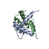
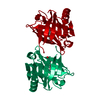
 Components
 Keywords
 Function and homology information
 Authors
 Poland, 1items
  Citation
 Structure visualization
 Downloads & links
 Other downloads


 PDBj
PDBj

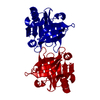
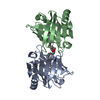
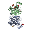
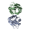
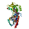
 Assembly
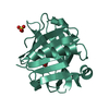


 Mass: 92.094 Da / Num. of mol.: 2 / Source method: obtained synthetically / Formula: C3H8O3
 Mass: 96.063 Da / Num. of mol.: 1 / Source method: obtained synthetically / Formula: SO4
 Mass: 18.015 Da / Num. of mol.: 59 / Source method: isolated from a natural source / Formula: H2O
 Sample preparation
 Processing