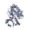
[English] 日本語
Yorodumi
- PDB-7b56: Crystal structure of CaMKII-actinin complex bound to AMPPNP -

+
Open data


ID or keywords:

Loading...

-
Basic information

Entry
Database: PDB / ID: 7b56
TitleCrystal structure of CaMKII-actinin complex bound to AMPPNP
Components
  • Alpha-actinin-2
  • Calcium/calmodulin-dependent protein kinase type II subunit alpha
KeywordsSTRUCTURAL PROTEIN / CaMKII / actinin / dendritic spine
Function / homology
Function and homology information


regulation of synaptic vesicle docking / glutamatergic postsynaptic density / actin filament uncapping / FATZ binding / titin Z domain binding / HSF1-dependent transactivation / Interferon gamma signaling / Ion transport by P-type ATPases / phospholipase C-activating angiotensin-activated signaling pathway / positive regulation of endocytic recycling ...regulation of synaptic vesicle docking / glutamatergic postsynaptic density / actin filament uncapping / FATZ binding / titin Z domain binding / HSF1-dependent transactivation / Interferon gamma signaling / Ion transport by P-type ATPases / phospholipase C-activating angiotensin-activated signaling pathway / positive regulation of endocytic recycling / Unblocking of NMDA receptors, glutamate binding and activation / peptidyl-threonine autophosphorylation / positive regulation of potassium ion transmembrane transporter activity / regulation of endocannabinoid signaling pathway / negative regulation of potassium ion transmembrane transporter activity / Trafficking of AMPA receptors / calcium- and calmodulin-dependent protein kinase complex / RAF activation / calmodulin dependent kinase signaling pathway / Ca2+ pathway / RAF/MAP kinase cascade / calcium-dependent protein serine/threonine kinase activity / NMDA selective glutamate receptor signaling pathway / regulation of neuron migration / Ca2+/calmodulin-dependent protein kinase / positive regulation of cation channel activity / LIM domain binding / negative regulation of protein localization to cell surface / regulation of neurotransmitter secretion / microspike assembly / postsynaptic actin cytoskeleton / dendritic spine development / muscle cell development / positive regulation of potassium ion transport / Ion homeostasis / positive regulation of calcium ion transport / postsynaptic specialization membrane / postsynaptic neurotransmitter receptor diffusion trapping / focal adhesion assembly / presynaptic cytosol / negative regulation of hydrolase activity / Assembly and cell surface presentation of NMDA receptors / Striated Muscle Contraction / calmodulin-dependent protein kinase activity / cardiac muscle cell development / Nephrin family interactions / GTPase activating protein binding / dendrite morphogenesis / regulation of mitochondrial membrane permeability involved in apoptotic process / sarcomere organization / regulation of neurotransmitter receptor localization to postsynaptic specialization membrane / postsynaptic cytosol / structural constituent of muscle / cortical actin cytoskeleton / Negative regulation of NMDA receptor-mediated neuronal transmission / positive regulation of cardiac muscle cell apoptotic process / Unblocking of NMDA receptors, glutamate binding and activation / regulation of neuronal synaptic plasticity / pseudopodium / postsynaptic density, intracellular component / negative regulation of potassium ion transport / Long-term potentiation / glutamate receptor binding / cellular response to interferon-beta / regulation of protein localization to plasma membrane / titin binding / phosphatidylinositol-4,5-bisphosphate binding / ionotropic glutamate receptor signaling pathway / regulation of membrane potential / Ras activation upon Ca2+ influx through NMDA receptor / cytoskeletal protein binding / dendrite cytoplasm / nuclear receptor coactivator activity / platelet alpha granule lumen / filopodium / cell projection / response to ischemia / actin filament / protein localization to plasma membrane / angiotensin-activated signaling pathway / postsynaptic density membrane / G1/S transition of mitotic cell cycle / positive regulation of receptor signaling pathway via JAK-STAT / Schaffer collateral - CA1 synapse / Z disc / cellular response to type II interferon / calcium ion transport / actin filament binding / integrin binding / Platelet degranulation / cell junction / kinase activity / actin cytoskeleton organization / RAF/MAP kinase cascade / peptidyl-serine phosphorylation / regulation of apoptotic process / transmembrane transporter binding / dendritic spine / postsynaptic density / cytoskeleton
Similarity search - Function
Calcium/calmodulin-dependent protein kinase II, association-domain / Calcium/calmodulin dependent protein kinase II association domain / EF-hand, Ca insensitive / Ca2+ insensitive EF hand / Ca2+ insensitive EF hand / Spectrin repeat / Spectrin repeat / Spectrin/alpha-actinin / Actinin-type actin-binding domain signature 1. / Actinin-type actin-binding domain signature 2. ...Calcium/calmodulin-dependent protein kinase II, association-domain / Calcium/calmodulin dependent protein kinase II association domain / EF-hand, Ca insensitive / Ca2+ insensitive EF hand / Ca2+ insensitive EF hand / Spectrin repeat / Spectrin repeat / Spectrin/alpha-actinin / Actinin-type actin-binding domain signature 1. / Actinin-type actin-binding domain signature 2. / Spectrin repeats / Actinin-type actin-binding domain, conserved site / Calponin homology domain / Calponin homology (CH) domain / Calponin homology domain / CH domain superfamily / Calponin homology (CH) domain profile. / NTF2-like domain superfamily / EF-hand domain pair / EF-hand, calcium binding motif / EF-hand calcium-binding domain profile. / EF-hand domain / EF-hand domain pair / Serine/threonine-protein kinase, active site / Serine/Threonine protein kinases active-site signature. / Protein kinase domain / Serine/Threonine protein kinases, catalytic domain / Protein kinase, ATP binding site / Protein kinases ATP-binding region signature. / Protein kinase domain profile. / Protein kinase domain / Protein kinase-like domain superfamily
Similarity search - Domain/homology
PHOSPHOAMINOPHOSPHONIC ACID-ADENYLATE ESTER / Calcium/calmodulin-dependent protein kinase type II subunit alpha / Alpha-actinin-2
Similarity search - Component
Biological speciesMus musculus (house mouse)
Homo sapiens (human)
MethodX-RAY DIFFRACTION / SYNCHROTRON / MOLECULAR REPLACEMENT / Resolution: 1.45 Å
AuthorsZhu, J. / Gold, M.
CitationJournal: To Be Published
Title: Crystal structure of CaMKII-actinin complex bound to MES
Authors: Zhu, J. / Gold, M.
History
DepositionDec 3, 2020Deposition site: PDBE / Processing site: PDBE
Revision 1.0Jun 22, 2022Provider: repository / Type: Initial release
Revision 1.1Jan 31, 2024Group: Data collection / Refinement description
Category: chem_comp_atom / chem_comp_bond / pdbx_initial_refinement_model

-
Structure visualization

Structure viewerMolecule:
MolmilJmol/JSmol

Downloads & links

-
Assembly

Deposited unit
B: Calcium/calmodulin-dependent protein kinase type II subunit alpha
A: Alpha-actinin-2
hetero molecules


Theoretical massNumber of molelcules
Total (without water)44,5335
Polymers43,8072
Non-polymers7263
Water7,945441
1


  • Idetical with deposited unit
  • defined by author&software
  • Evidence: gel filtration
TypeNameSymmetry operationNumber
identity operation1_555x,y,z1
Buried area3470 Å2
ΔGint-21 kcal/mol
Surface area16560 Å2
MethodPISA
Unit cell
Length a, b, c (Å)64.426, 72.510, 92.001
Angle α, β, γ (deg.)90.000, 90.000, 90.000
Int Tables number19
Space group name H-MP212121
Space group name HallP2ac2ab
Symmetry operation#1: x,y,z
#2: x+1/2,-y+1/2,-z
#3: -x,y+1/2,-z+1/2
#4: -x+1/2,-y,z+1/2

-
Components

-
Protein , 2 types, 2 molecules BA

#1: Protein Calcium/calmodulin-dependent protein kinase type II subunit alpha / / CaMK-II subunit alpha


Mass: 35903.430 Da / Num. of mol.: 1
Source method: isolated from a genetically manipulated source
Source: (gene. exp.) Mus musculus (house mouse) / Gene: Camk2a / Production host: Escherichia coli (E. coli)
References: UniProt: P11798, Ca2+/calmodulin-dependent protein kinase
#2: Protein Alpha-actinin-2 / / Alpha-actinin skeletal muscle isoform 2 / F-actin cross-linking protein


Mass: 7903.778 Da / Num. of mol.: 1
Source method: isolated from a genetically manipulated source
Source: (gene. exp.) Homo sapiens (human) / Gene: ACTN2 / Production host: Escherichia coli (E. coli) / References: UniProt: P35609

-
Non-polymers , 4 types, 444 molecules

#3: Chemical ChemComp-ANP / PHOSPHOAMINOPHOSPHONIC ACID-ADENYLATE ESTER


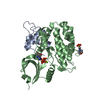
Mass: 506.196 Da / Num. of mol.: 1 / Source method: obtained synthetically / Formula: C10H17N6O12P3 / Feature type: SUBJECT OF INVESTIGATION / Comment: AMP-PNP, energy-carrying molecule analogue*YM
#4: Chemical ChemComp-MG / MAGNESIUM ION


Mass: 24.305 Da / Num. of mol.: 1 / Source method: obtained synthetically / Formula: Mg / Feature type: SUBJECT OF INVESTIGATION
#5: Chemical ChemComp-MES / 2-(N-MORPHOLINO)-ETHANESULFONIC ACID / MES (buffer)


Mass: 195.237 Da / Num. of mol.: 1 / Source method: obtained synthetically / Formula: C6H13NO4S / Feature type: SUBJECT OF INVESTIGATION / Comment: pH buffer*YM
#6: Water ChemComp-HOH / water / Water


Mass: 18.015 Da / Num. of mol.: 441 / Source method: isolated from a natural source / Formula: H2O

-
Details

Has ligand of interestY

-
Experimental details

-
Experiment

ExperimentMethod: X-RAY DIFFRACTION / Number of used crystals: 1

-
Sample preparation

CrystalDensity Matthews: 2.45 Å3/Da / Density % sol: 49.85 %
Crystal growTemperature: 277 K / Method: vapor diffusion, sitting drop
Details: 0.1 M MES pH 6.0, 20% w/v PEG4000, 0.2 M Lithium Sulfate

-
Data collection

DiffractionMean temperature: 100 K / Serial crystal experiment: N
Diffraction sourceSource: SYNCHROTRON / Site: Diamond / Beamline: I04-1 / Wavelength: 0.9119 Å
DetectorType: DECTRIS PILATUS3 S 6M / Detector: PIXEL / Date: Mar 1, 2020
RadiationProtocol: SINGLE WAVELENGTH / Monochromatic (M) / Laue (L): M / Scattering type: x-ray
Radiation wavelengthWavelength: 0.9119 Å / Relative weight: 1
ReflectionResolution: 1.45→92 Å / Num. obs: 77065 / % possible obs: 100 % / Redundancy: 12.1 % / Biso Wilson estimate: 22.55 Å2 / CC1/2: 0.999 / Rpim(I) all: 0.016 / Net I/σ(I): 18.5
Reflection shellResolution: 1.45→1.47 Å / Num. unique obs: 3786 / CC1/2: 0.825 / Rpim(I) all: 0.522

-
Processing

Software
NameVersionClassification
PHENIX1.18.2_3874refinement
DIALSdata reduction
Aimlessdata scaling
MOLREPphasing
RefinementMethod to determine structure: MOLECULAR REPLACEMENT
Starting model: 2VZ6
Resolution: 1.45→52.77 Å / SU ML: 0.1456 / Cross valid method: FREE R-VALUE / σ(F): 1.34 / Phase error: 22.2221
Stereochemistry target values: GeoStd + Monomer Library + CDL v1.2
RfactorNum. reflection% reflection
Rfree0.2154 1996 2.6 %
Rwork0.1901 74889 -
obs0.1908 76885 99.83 %
Solvent computationShrinkage radii: 0.9 Å / VDW probe radii: 1.11 Å / Solvent model: FLAT BULK SOLVENT MODEL
Displacement parametersBiso mean: 30.22 Å2
Refinement stepCycle: LAST / Resolution: 1.45→52.77 Å
ProteinNucleic acidLigandSolventTotal
Num. atoms2900 0 44 441 3385
Refine LS restraints
Refine-IDTypeDev idealNumber
X-RAY DIFFRACTIONf_bond_d0.00573049
X-RAY DIFFRACTIONf_angle_d0.97424149
X-RAY DIFFRACTIONf_chiral_restr0.0763456
X-RAY DIFFRACTIONf_plane_restr0.0058529
X-RAY DIFFRACTIONf_dihedral_angle_d25.08671121
LS refinement shell
Resolution (Å)Rfactor RfreeNum. reflection RfreeRfactor RworkNum. reflection RworkRefine-ID% reflection obs (%)
1.45-1.490.28411400.28455283X-RAY DIFFRACTION99.69
1.49-1.530.23621400.24945259X-RAY DIFFRACTION99.7
1.53-1.570.23951420.2265300X-RAY DIFFRACTION99.67
1.57-1.620.24291400.21135259X-RAY DIFFRACTION99.58
1.62-1.680.23151410.20595292X-RAY DIFFRACTION99.72
1.68-1.750.26571420.21665313X-RAY DIFFRACTION99.73
1.75-1.830.21811410.20265302X-RAY DIFFRACTION99.83
1.83-1.920.23651420.19655294X-RAY DIFFRACTION99.89
1.92-2.040.20961420.19235356X-RAY DIFFRACTION99.91
2.04-2.20.22751430.19395359X-RAY DIFFRACTION100
2.2-2.420.18491430.19085374X-RAY DIFFRACTION100
2.42-2.770.25631440.20125398X-RAY DIFFRACTION99.96
2.77-3.490.20671450.18695436X-RAY DIFFRACTION100
3.49-52.770.19971510.17175664X-RAY DIFFRACTION99.97
Refinement TLS params.Method: refined / Origin x: 15.1121581365 Å / Origin y: -5.04463431322 Å / Origin z: -2.5044161769 Å
111213212223313233
T0.155220329519 Å2-0.00909408337564 Å20.0156327723026 Å2-0.17085218412 Å2-0.01153362473 Å2--0.1753018623 Å2
L0.4910913964 °2-0.044831707403 °20.236917221921 °2-0.406183786294 °2-0.149582352756 °2--0.728566639229 °2
S-0.00983201735945 Å °0.0262968054646 Å °-0.0150357802084 Å °0.0277117250096 Å °0.0342016859214 Å °-0.0299469579307 Å °-0.0709124387588 Å °0.103534743467 Å °-1.39634220456E-6 Å °
Refinement TLS groupSelection details: all

+
About Yorodumi

-
News

-
Feb 9, 2022. New format data for meta-information of EMDB entries

New format data for meta-information of EMDB entries

  • Version 3 of the EMDB header file is now the official format.
  • The previous official version 1.9 will be removed from the archive.

Related info.:EMDB header

External links:wwPDB to switch to version 3 of the EMDB data model

-
Aug 12, 2020. Covid-19 info

Covid-19 info

URL: https://pdbj.org/emnavi/covid19.php

New page: Covid-19 featured information page in EM Navigator.

Related info.:Covid-19 info / Mar 5, 2020. Novel coronavirus structure data

+
Mar 5, 2020. Novel coronavirus structure data

Novel coronavirus structure data

Related info.:Yorodumi Speices / Aug 12, 2020. Covid-19 info

External links:COVID-19 featured content - PDBj / Molecule of the Month (242):Coronavirus Proteases

+
Jan 31, 2019. EMDB accession codes are about to change! (news from PDBe EMDB page)

EMDB accession codes are about to change! (news from PDBe EMDB page)

  • The allocation of 4 digits for EMDB accession codes will soon come to an end. Whilst these codes will remain in use, new EMDB accession codes will include an additional digit and will expand incrementally as the available range of codes is exhausted. The current 4-digit format prefixed with “EMD-” (i.e. EMD-XXXX) will advance to a 5-digit format (i.e. EMD-XXXXX), and so on. It is currently estimated that the 4-digit codes will be depleted around Spring 2019, at which point the 5-digit format will come into force.
  • The EM Navigator/Yorodumi systems omit the EMD- prefix.

Related info.:Q: What is EMD? / ID/Accession-code notation in Yorodumi/EM Navigator

External links:EMDB Accession Codes are Changing Soon! / Contact to PDBj

+
Jul 12, 2017. Major update of PDB

Major update of PDB

  • wwPDB released updated PDB data conforming to the new PDBx/mmCIF dictionary.
  • This is a major update changing the version number from 4 to 5, and with Remediation, in which all the entries are updated.
  • In this update, many items about electron microscopy experimental information are reorganized (e.g. em_software).
  • Now, EM Navigator and Yorodumi are based on the updated data.

External links:wwPDB Remediation / Enriched Model Files Conforming to OneDep Data Standards Now Available in the PDB FTP Archive

-
Yorodumi

Thousand views of thousand structures

  • Yorodumi is a browser for structure data from EMDB, PDB, SASBDB, etc.
  • This page is also the successor to EM Navigator detail page, and also detail information page/front-end page for Omokage search.
  • The word "yorodu" (or yorozu) is an old Japanese word meaning "ten thousand". "mi" (miru) is to see.

Related info.:EMDB / PDB / SASBDB / Comparison of 3 databanks / Yorodumi Search / Aug 31, 2016. New EM Navigator & Yorodumi / Yorodumi Papers / Jmol/JSmol / Function and homology information / Changes in new EM Navigator and Yorodumi

Read more