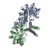
 Movie
Movie Controller

[English] 日本語
 Yorodumi
- PDB-7b1r: Crystal structure of B. subtilis glucose-1-phosphate uridylyltran... -
+ Open data

- Basic information
| Entry | Database: PDB / ID: 7b1r | ||||||
|---|---|---|---|---|---|---|---|
| Title | Crystal structure of B. subtilis glucose-1-phosphate uridylyltransferase YngB | ||||||
 Components Components | Probable UTP--glucose-1-phosphate uridylyltransferase YngB | ||||||
 Keywords Keywords |  TRANSFERASE / UDP-glucose / glucose-1-phosphate / UTP TRANSFERASE / UDP-glucose / glucose-1-phosphate / UTP | ||||||
| Function / homology | UTP--glucose-1-phosphate uridylyltransferase, bacterial/archaeal-type / enterobacterial common antigen biosynthetic process / UTP-glucose-1-phosphate uridylyltransferase / UTP:glucose-1-phosphate uridylyltransferase activity / UDP-glucose metabolic process / Nucleotidyl transferase domain / Nucleotidyl transferase / Nucleotide-diphospho-sugar transferases / UTP--glucose-1-phosphate uridylyltransferase YngB Function and homology information Function and homology information | ||||||
| Biological species |  Bacillus subtilis subsp. subtilis str. 168 (bacteria) Bacillus subtilis subsp. subtilis str. 168 (bacteria) | ||||||
| Method | X-RAY DIFFRACTION / SYNCHROTRON / SAD / Resolution: 2.8 Å | ||||||
 Authors Authors | Wu, C. / Morgan, R.M.L. / Freemont, P. / Grundling, A. | ||||||
| Funding support |  United Kingdom, 1items United Kingdom, 1items
| ||||||
 Citation Citation | Journal: J.Biol.Chem. / Year: 2021 Title: Bacillus subtilis YngB contributes to wall teichoic acid glucosylation and glycolipid formation during anaerobic growth. Authors: Wu, C.H. / Rismondo, J. / Morgan, R.M.L. / Shen, Y. / Loessner, M.J. / Larrouy-Maumus, G. / Freemont, P.S. / Grundling, A. | ||||||
| History |
|
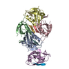
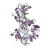
- Structure visualization
| Structure viewer | Molecule: MolmilJmol/JSmol |
|---|
- Downloads & links
-Download
| PDBx/mmCIF format | 7b1r.cif.gz | 122.2 KB | Display | PDBx/mmCIF format |
|---|---|---|---|---|
| PDB format | pdb7b1r.ent.gz | 98.7 KB | Display | PDB format |
| PDBx/mmJSON format | 7b1r.json.gz | Tree view | PDBx/mmJSON format | |
| Others |  Other downloads Other downloads |
-Validation report
| Arichive directory | https://data.pdbj.org/pub/pdb/validation_reports/b1/7b1rftp://data.pdbj.org/pub/pdb/validation_reports/b1/7b1r | HTTPS FTP |
|---|
-Related structure data
| Similar structure data |
|---|


-Links
 PDBj
PDBj

- Assembly
| Deposited unit | 
| ||||||||
|---|---|---|---|---|---|---|---|---|---|
| 1 | 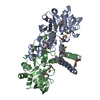
| ||||||||
| Unit cell |
| ||||||||
| Components on special symmetry positions |
|
-Components
| #1: Protein | Mass: 33548.262 Da / Num. of mol.: 2 Source method: isolated from a genetically manipulated source Source: (gene. exp.) Bacillus subtilis subsp. subtilis str. 168 (bacteria)Gene: yngB, BSU18180 / Production host: Escherichia coli (E. coli)References: UniProt: O31822, UTP-glucose-1-phosphate uridylyltransferase#2: Water | ChemComp-HOH / | Water Mass: 18.015 Da / Num. of mol.: 54 / Source method: isolated from a natural source / Formula: H2O Mass: 18.015 Da / Num. of mol.: 54 / Source method: isolated from a natural source / Formula: H2OHas ligand of interest | N | |
|---|
-Experimental details
-Experiment
| Experiment | Method: X-RAY DIFFRACTION / Number of used crystals: 1 |
|---|
- Sample preparation
| Crystal | Density Matthews: 2.94 Å3/Da / Density % sol: 58.14 % |
|---|---|
| Crystal grow | Temperature: 289 K / Method: vapor diffusion, sitting drop Details: 0.2M potassium citrate tribasic monohydrate, 0.05M lithium citrate tribasic tetrahydrate, 0.1M sodium phosphate monobasic monohydrate, 25% PEG6000 |
-Data collection
| Diffraction | Mean temperature: 100 K / Serial crystal experiment: N |
|---|---|
| Diffraction source | Source: SYNCHROTRON / Site: Diamond / Beamline: I03 / Wavelength: 0.9794 Å |
| Detector | Type: DECTRIS EIGER2 XE 16M / Detector: PIXEL / Date: Feb 5, 2020 |
| Radiation | Protocol: SINGLE WAVELENGTH / Monochromatic (M) / Laue (L): M / Scattering type: x-ray |
| Radiation wavelength | Wavelength: 0.9794 Å / Relative weight: 1 |
| Reflection | Resolution: 2.8→55.98 Å / Num. obs: 19457 / % possible obs: 99.8 % / Redundancy: 1.9 % / Rpim(I) all: 0.067 / Rrim(I) all: 0.095 / Net I/σ(I): 5.6 |
| Reflection shell | Resolution: 2.8→2.95 Å / Num. unique obs: 2777 / Rpim(I) all: 0.468 / Rrim(I) all: 0.661 |
-Phasing
| Phasing | Method: SAD |
|---|
- Processing
| Software |
| |||||||||||||||||||||||||||||||||||||||||||||
|---|---|---|---|---|---|---|---|---|---|---|---|---|---|---|---|---|---|---|---|---|---|---|---|---|---|---|---|---|---|---|---|---|---|---|---|---|---|---|---|---|---|---|---|---|---|---|
| Refinement | Method to determine structure: SAD / Resolution: 2.8→51.09 Å / Cor.coef. Fo:Fc: 0.959 / Cor.coef. Fo:Fc free: 0.942 / SU B: 16.135 / SU ML: 0.296 / Cross valid method: THROUGHOUT / σ(F): 0 / ESU R: 1.385 / ESU R Free: 0.342 / Stereochemistry target values: MAXIMUM LIKELIHOOD / Details: U VALUES : REFINED INDIVIDUALLY
| |||||||||||||||||||||||||||||||||||||||||||||
| Solvent computation | Ion probe radii: 0.8 Å / Shrinkage radii: 0.8 Å / VDW probe radii: 1.2 Å / Solvent model: MASK | |||||||||||||||||||||||||||||||||||||||||||||
| Displacement parameters | Biso max: 256.86 Å2 / Biso mean: 82.119 Å2 / Biso min: 42.19 Å2
| |||||||||||||||||||||||||||||||||||||||||||||
| Refinement step | Cycle: final / Resolution: 2.8→51.09 Å
| |||||||||||||||||||||||||||||||||||||||||||||
| Refine LS restraints |
| |||||||||||||||||||||||||||||||||||||||||||||
| LS refinement shell | Resolution: 2.8→2.873 Å / Rfactor Rfree error: 0 / Total num. of bins used: 20
|
