 Movie
Movie Controller

[English] 日本語
 Yorodumi
- PDB-7axp: Structural characterisation of WDR5:CS-VIP8 interaction in cis state 2 -
+ Open data

- Basic information
| Entry | Database: PDB / ID: 7axp | |||||||||
|---|---|---|---|---|---|---|---|---|---|---|
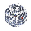
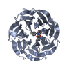
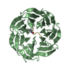
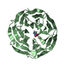
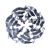
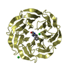
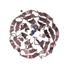
| Title | Structural characterisation of WDR5:CS-VIP8 interaction in cis state 2 | |||||||||
 Components Components |
| |||||||||
 Keywords Keywords |  TRANSFERASE / WDR5 / cyclic strained visible-light photoswitches / MLL1 complex disruption / inhibition of hematopoiesis TRANSFERASE / WDR5 / cyclic strained visible-light photoswitches / MLL1 complex disruption / inhibition of hematopoiesis | |||||||||
| Function / homology |  Function and homology information Function and homology informationMLL3/4 complex / Set1C/COMPASS complex / MLL1/2 complex / ATAC complex / NSL complex / histone H3K4 methyltransferase activity / : / Cardiogenesis / histone methyltransferase complex / regulation of tubulin deacetylation ...MLL3/4 complex / Set1C/COMPASS complex / MLL1/2 complex / ATAC complex / NSL complex / histone H3K4 methyltransferase activity / : / Cardiogenesis / histone methyltransferase complex / regulation of tubulin deacetylation / Formation of WDR5-containing histone-modifying complexes / regulation of cell division / MLL1 complex / regulation of embryonic development / histone acetyltransferase complex / positive regulation of gluconeogenesis / transcription initiation-coupled chromatin remodeling / methylated histone binding / skeletal system development / gluconeogenesis / RUNX1 regulates genes involved in megakaryocyte differentiation and platelet function / mitotic spindle / PKMTs methylate histone lysines / RMTs methylate histone arginines / Activation of anterior HOX genes in hindbrain development during early embryogenesis / Neddylation / HATs acetylate histones / histone binding / regulation of cell cycle / regulation of DNA-templated transcription / regulation of transcription by RNA polymerase II / positive regulation of DNA-templated transcription / negative regulation of transcription by RNA polymerase II / nucleoplasm / nucleusSimilarity search - Function | |||||||||
| Biological species |  Homo sapiens (human) Homo sapiens (human) | |||||||||
| Method | X-RAY DIFFRACTION / SYNCHROTRON / MOLECULAR REPLACEMENT / Resolution: 2.432 Å | |||||||||
 Authors Authors | Werel, L. / Essen, L.-O. | |||||||||
 Citation Citation | Journal: Acs Cent.Sci. / Year: 2022 Title: Bistable Photoswitch Allows in Vivo Control of Hematopoiesis. Authors: Albert, L. / Nagpal, J. / Steinchen, W. / Zhang, L. / Werel, L. / Djokovic, N. / Ruzic, D. / Hoffarth, M. / Xu, J. / Kaspareit, J. / Abendroth, F. / Royant, A. / Bange, G. / Nikolic, K. / ...Authors: Albert, L. / Nagpal, J. / Steinchen, W. / Zhang, L. / Werel, L. / Djokovic, N. / Ruzic, D. / Hoffarth, M. / Xu, J. / Kaspareit, J. / Abendroth, F. / Royant, A. / Bange, G. / Nikolic, K. / Ryu, S. / Dou, Y. / Essen, L.O. / Vazquez, O. | |||||||||
| History |
|
- Structure visualization
| Structure viewer | Molecule: MolmilJmol/JSmol |
|---|
- Downloads & links
-Download
| PDBx/mmCIF format | 7axp.cif.gz | 136.6 KB | Display | PDBx/mmCIF format |
|---|---|---|---|---|
| PDB format | pdb7axp.ent.gz | 106.4 KB | Display | PDB format |
| PDBx/mmJSON format | 7axp.json.gz | Tree view | PDBx/mmJSON format | |
| Others |  Other downloads Other downloads |
-Validation report
| Arichive directory | https://data.pdbj.org/pub/pdb/validation_reports/ax/7axpftp://data.pdbj.org/pub/pdb/validation_reports/ax/7axp | HTTPS FTP |
|---|
-Related structure data
| Related structure data |  7axqC  7axsC  7axuC  7axxC  6iamS S: Starting model for refinement C: citing same article ( |
|---|---|
| Similar structure data |

-Links
 PDBj
PDBj- Assembly
| Deposited unit | 
| ||||||||
|---|---|---|---|---|---|---|---|---|---|
| 1 |
| ||||||||
| Unit cell |
|
-Components
| #1: Protein | Mass: 36635.438 Da / Num. of mol.: 1 Source method: isolated from a genetically manipulated source Details: WDR5, component of MLL1 methyltransferase complex, chromatin regulator Source: (gene. exp.) Homo sapiens (human) / Gene: WDR5, BIG3 / Production host:  Escherichia coli BL21 (bacteria) / References: UniProt: P61964 Escherichia coli BL21 (bacteria) / References: UniProt: P61964 |
|---|---|
| #2: Protein/peptide | Mass: 915.999 Da / Num. of mol.: 1 / Source method: obtained synthetically Details: cyclic peptide, cyclic strained visible-light photoswitch, WDR5-binder, disrupts MLL1 complex via MLL1-WDR5 inhibition Source: (synth.) Homo sapiens (human) |
| #3: Water | ChemComp-HOH / Water Mass: 18.015 Da / Num. of mol.: 20 / Source method: isolated from a natural source / Formula: H2O Mass: 18.015 Da / Num. of mol.: 20 / Source method: isolated from a natural source / Formula: H2O |
| Has ligand of interest | N |
-Experimental details
-Experiment
| Experiment | Method: X-RAY DIFFRACTION / Number of used crystals: 1 |
|---|
- Sample preparation
| Crystal | Density Matthews: 3.76 Å3/Da / Density % sol: 67.3 % |
|---|---|
| Crystal grow | Temperature: 277.15 K / Method: vapor diffusion, sitting drop / pH: 8.5 Details: 10% (w/v) PEG20000, 20% (v/v) PEG550 MME, 0.02 M sodium formate, 0.02 M ammonium acetate, 0.02 M trisodium citrate, 0.02 M sodium potassium L-tartrate, 0.02 M sodium oxamate |
-Data collection
| Diffraction | Mean temperature: 100 K / Serial crystal experiment: N |
|---|---|
| Diffraction source | Source: SYNCHROTRON / Site: PETRA III, EMBL c/o DESY  / Beamline: P13 (MX1) / Wavelength: 1 Å / Beamline: P13 (MX1) / Wavelength: 1 Å |
| Detector | Type: DECTRIS PILATUS 6M / Detector: PIXEL / Date: Mar 30, 2018 |
| Radiation | Protocol: SINGLE WAVELENGTH / Monochromatic (M) / Laue (L): M / Scattering type: x-ray |
| Radiation wavelength | Wavelength: 1 Å / Relative weight: 1 |
| Reflection | Resolution: 2.432→64.765 Å / Num. obs: 155702 / % possible obs: 99.7 % / Redundancy: 13.6 % / CC1/2: 0.996 / Rmerge(I) obs: 0.086 / Net I/σ(I): 16.5 |
| Reflection shell | Resolution: 2.432→2.519 Å / Redundancy: 12.8 % / Num. unique obs: 92 / CC1/2: 0.995 / % possible all: 0.998 |
- Processing
| Software |
| |||||||||||||||||||||||||||||||||||||||||||||||||||||||||||||||||||||||||||||||||||||||||||||||||||||||||||||||||||||||||||||
|---|---|---|---|---|---|---|---|---|---|---|---|---|---|---|---|---|---|---|---|---|---|---|---|---|---|---|---|---|---|---|---|---|---|---|---|---|---|---|---|---|---|---|---|---|---|---|---|---|---|---|---|---|---|---|---|---|---|---|---|---|---|---|---|---|---|---|---|---|---|---|---|---|---|---|---|---|---|---|---|---|---|---|---|---|---|---|---|---|---|---|---|---|---|---|---|---|---|---|---|---|---|---|---|---|---|---|---|---|---|---|---|---|---|---|---|---|---|---|---|---|---|---|---|---|---|---|
| Refinement | Method to determine structure: MOLECULAR REPLACEMENT Starting model: 6IAM Resolution: 2.432→64.765 Å / SU ML: 0.33 / Cross valid method: THROUGHOUT / σ(F): 1.35 / Phase error: 44.28 / Stereochemistry target values: ML
| |||||||||||||||||||||||||||||||||||||||||||||||||||||||||||||||||||||||||||||||||||||||||||||||||||||||||||||||||||||||||||||
| Solvent computation | Shrinkage radii: 0.9 Å / VDW probe radii: 1.11 Å / Solvent model: FLAT BULK SOLVENT MODEL | |||||||||||||||||||||||||||||||||||||||||||||||||||||||||||||||||||||||||||||||||||||||||||||||||||||||||||||||||||||||||||||
| Displacement parameters | Biso max: 165.08 Å2 / Biso mean: 67.6577 Å2 / Biso min: 20.47 Å2 | |||||||||||||||||||||||||||||||||||||||||||||||||||||||||||||||||||||||||||||||||||||||||||||||||||||||||||||||||||||||||||||
| Refinement step | Cycle: final / Resolution: 2.432→64.765 Å
| |||||||||||||||||||||||||||||||||||||||||||||||||||||||||||||||||||||||||||||||||||||||||||||||||||||||||||||||||||||||||||||
| LS refinement shell | Refine-ID: X-RAY DIFFRACTION / Rfactor Rfree error: 0
| |||||||||||||||||||||||||||||||||||||||||||||||||||||||||||||||||||||||||||||||||||||||||||||||||||||||||||||||||||||||||||||
| Refinement TLS params. | Method: refined / Refine-ID: X-RAY DIFFRACTION
| |||||||||||||||||||||||||||||||||||||||||||||||||||||||||||||||||||||||||||||||||||||||||||||||||||||||||||||||||||||||||||||
| Refinement TLS group |
|
