 Movie
Movie Controller

[English] 日本語
 Yorodumi
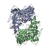
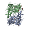
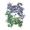
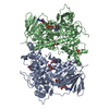
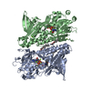
- PDB-7aqu: Flavin-dependent tryptophan halogenase Thal: N-terminally His-tag... -
+ Open data

- Basic information
| Entry | Database: PDB / ID: 7aqu | ||||||
|---|---|---|---|---|---|---|---|
| Title | Flavin-dependent tryptophan halogenase Thal: N-terminally His-tagged form of quintuple mutant (NHis-Thal-RebH5) | ||||||
 Components Components | Tryptophan 6-halogenase | ||||||
 Keywords Keywords |  FLAVOPROTEIN / tryptophan halogenase / ThdH / His-Tag FLAVOPROTEIN / tryptophan halogenase / ThdH / His-Tag | ||||||
| Function / homology |  Function and homology information Function and homology information | ||||||
| Biological species |  Streptomyces albogriseolus (bacteria) Streptomyces albogriseolus (bacteria) | ||||||
| Method | X-RAY DIFFRACTION / SYNCHROTRON / MOLECULAR REPLACEMENT / Resolution: 1.63 Å | ||||||
 Authors Authors | Moritzer, A.C. / Prior, T. / Niemann, H.H. | ||||||
 Citation Citation | Journal: Crystals / Year: 2020 Title: Not Cleaving the His-tag of Thal Results in More Tightly Packed and Better-Diffracting Crystals Authors: Moritzer, A.C. / Prior, T. / Niemann, H.H. | ||||||
| History |
|
- Structure visualization
| Structure viewer | Molecule: MolmilJmol/JSmol |
|---|
- Downloads & links
-Download
| PDBx/mmCIF format | 7aqu.cif.gz | 257.7 KB | Display | PDBx/mmCIF format |
|---|---|---|---|---|
| PDB format | pdb7aqu.ent.gz | 197.6 KB | Display | PDB format |
| PDBx/mmJSON format | 7aqu.json.gz | Tree view | PDBx/mmJSON format | |
| Others |  Other downloads Other downloads |
-Validation report
| Arichive directory | https://data.pdbj.org/pub/pdb/validation_reports/aq/7aquftp://data.pdbj.org/pub/pdb/validation_reports/aq/7aqu | HTTPS FTP |
|---|
-Related structure data
| Related structure data |  7aqvC  6h43S  6ylw 6z6t S: Starting model for refinement C: citing same article ( |
|---|---|
| Similar structure data |


-Links
 PDBj
PDBj

- Assembly
| Deposited unit | 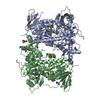
| ||||||||||||||||||
|---|---|---|---|---|---|---|---|---|---|---|---|---|---|---|---|---|---|---|---|
| 1 |
| ||||||||||||||||||
| Unit cell |
| ||||||||||||||||||
| Noncrystallographic symmetry (NCS) | NCS domain:
NCS domain segments: Component-ID: 0 / Ens-ID: 1 / Beg auth comp-ID: ASP / Beg label comp-ID: ASP / End auth comp-ID: GLY / End label comp-ID: GLY / Refine code: 0 / Auth seq-ID: 2 - 529 / Label seq-ID: 22 - 549
|
-Components
-Protein , 1 types, 2 molecules AB
| #1: Protein | Mass: 62362.312 Da / Num. of mol.: 2 / Mutation: V52I, V82I, S360T, G469S, S470N Source method: isolated from a genetically manipulated source Details: N-terminal His-tag from pET-28a not cleaved / Source: (gene. exp.) Streptomyces albogriseolus (bacteria) / Gene: thal, thdH / Plasmid: pET-28a / Production host: Escherichia coli BL21(DE3) (bacteria) / References: UniProt: A1E280 |
|---|
-Non-polymers , 6 types, 990 molecules 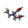


| #2: Chemical | ChemComp-BCN / Bicine Mass: 163.172 Da / Num. of mol.: 1 / Source method: obtained synthetically / Formula: C6H13NO4 / Comment: pH buffer*YM Mass: 163.172 Da / Num. of mol.: 1 / Source method: obtained synthetically / Formula: C6H13NO4 / Comment: pH buffer*YM | ||||||||
|---|---|---|---|---|---|---|---|---|---|
| #3: Chemical | ChemComp-GOL / Glycerol Mass: 92.094 Da / Num. of mol.: 5 / Source method: obtained synthetically / Formula: C3H8O3 Mass: 92.094 Da / Num. of mol.: 5 / Source method: obtained synthetically / Formula: C3H8O3#4: Chemical | ChemComp-GLY / Glycine Type: peptide linking / Mass: 75.067 Da / Num. of mol.: 7 / Source method: obtained synthetically / Formula: C2H5NO2 Type: peptide linking / Mass: 75.067 Da / Num. of mol.: 7 / Source method: obtained synthetically / Formula: C2H5NO2#5: Chemical | ChemComp-ALA / | Alanine Type: L-peptide linking / Mass: 89.093 Da / Num. of mol.: 1 / Source method: obtained synthetically / Formula: C3H7NO2 Type: L-peptide linking / Mass: 89.093 Da / Num. of mol.: 1 / Source method: obtained synthetically / Formula: C3H7NO2#6: Chemical | ChemComp-SER / | Serine Type: L-peptide linking / Mass: 105.093 Da / Num. of mol.: 1 / Source method: obtained synthetically / Formula: C3H7NO3 Type: L-peptide linking / Mass: 105.093 Da / Num. of mol.: 1 / Source method: obtained synthetically / Formula: C3H7NO3#7: Water | ChemComp-HOH / | WaterMass: 18.015 Da / Num. of mol.: 975 / Source method: isolated from a natural source / Formula: H2O |
-Details
| Has ligand of interest | N |
|---|
-Experimental details
-Experiment
| Experiment | Method: X-RAY DIFFRACTION / Number of used crystals: 1 |
|---|
- Sample preparation
| Crystal | Density Matthews: 2.3 Å3/Da / Density % sol: 46.51 % / Description: Hexagonal |
|---|---|
| Crystal grow | Temperature: 293.5 K / Method: vapor diffusion, sitting drop / pH: 9 Details: reservoir solution: 0.1 M bicine pH 9.0, 20 % (w/v) PEG 4000, 10 % (v/v) glycerol, 0.02 M amino acid mix consisting of L-Glu, L-Ala, D-Ala, Gly, L-Lys, D-Lysis, L-Ser and D-Ser; protein ...Details: reservoir solution: 0.1 M bicine pH 9.0, 20 % (w/v) PEG 4000, 10 % (v/v) glycerol, 0.02 M amino acid mix consisting of L-Glu, L-Ala, D-Ala, Gly, L-Lys, D-Lysis, L-Ser and D-Ser; protein buffer solution: 10 mM TRIS, 50 mM NaCl, 1 mM TCEP; protein concentration: ~15 mg/mL; drop ratio: 100 nL+100 nL (protein + reservoir) |
-Data collection
| Diffraction | Mean temperature: 100 K / Serial crystal experiment: N |
|---|---|
| Diffraction source | Source: SYNCHROTRON / Site: PETRA III, EMBL c/o DESY  / Beamline: P13 (MX1) / Wavelength: 0.9763 Å / Beamline: P13 (MX1) / Wavelength: 0.9763 Å |
| Detector | Type: DECTRIS PILATUS 6M / Detector: PIXEL / Date: Jun 18, 2018 |
| Radiation | Monochromator: DCM / Protocol: SINGLE WAVELENGTH / Monochromatic (M) / Laue (L): M / Scattering type: x-ray |
| Radiation wavelength | Wavelength: 0.9763 Å / Relative weight: 1 |
| Reflection | Resolution: 1.63→50 Å / Num. obs: 129082 / % possible obs: 96.6 % / Redundancy: 7.03 % / Biso Wilson estimate: 31.4 Å2 / CC1/2: 0.999 / Rrim(I) all: 0.079 / Net I/σ(I): 15.33 |
| Reflection shell | Resolution: 1.63→1.67 Å / Redundancy: 6.73 % / Mean I/σ(I) obs: 1.97 / Num. unique obs: 8424 / CC1/2: 0.748 / Rrim(I) all: 1.146 / % possible all: 85.3 |
- Processing
| Software |
| ||||||||||||||||||||||||||||||||||||||||||||||||||||||||||||||||||||||||||||||||||||||||||||||||||||||||||||||||||||||||||||||||||||||||||||||||||||||||||||||||
|---|---|---|---|---|---|---|---|---|---|---|---|---|---|---|---|---|---|---|---|---|---|---|---|---|---|---|---|---|---|---|---|---|---|---|---|---|---|---|---|---|---|---|---|---|---|---|---|---|---|---|---|---|---|---|---|---|---|---|---|---|---|---|---|---|---|---|---|---|---|---|---|---|---|---|---|---|---|---|---|---|---|---|---|---|---|---|---|---|---|---|---|---|---|---|---|---|---|---|---|---|---|---|---|---|---|---|---|---|---|---|---|---|---|---|---|---|---|---|---|---|---|---|---|---|---|---|---|---|---|---|---|---|---|---|---|---|---|---|---|---|---|---|---|---|---|---|---|---|---|---|---|---|---|---|---|---|---|---|---|---|---|
| Refinement | Method to determine structure: MOLECULAR REPLACEMENT Starting model: 6H43 chain A Resolution: 1.63→48.579 Å / Cor.coef. Fo:Fc: 0.973 / Cor.coef. Fo:Fc free: 0.961 / SU B: 2.211 / SU ML: 0.073 / Cross valid method: FREE R-VALUE / ESU R: 0.095 / ESU R Free: 0.095 Details: Hydrogens have been added in their riding positions
| ||||||||||||||||||||||||||||||||||||||||||||||||||||||||||||||||||||||||||||||||||||||||||||||||||||||||||||||||||||||||||||||||||||||||||||||||||||||||||||||||
| Solvent computation | Ion probe radii: 0.8 Å / Shrinkage radii: 0.8 Å / VDW probe radii: 1.2 Å / Solvent model: MASK BULK SOLVENT | ||||||||||||||||||||||||||||||||||||||||||||||||||||||||||||||||||||||||||||||||||||||||||||||||||||||||||||||||||||||||||||||||||||||||||||||||||||||||||||||||
| Displacement parameters | Biso mean: 27.22 Å2
| ||||||||||||||||||||||||||||||||||||||||||||||||||||||||||||||||||||||||||||||||||||||||||||||||||||||||||||||||||||||||||||||||||||||||||||||||||||||||||||||||
| Refinement step | Cycle: LAST / Resolution: 1.63→48.579 Å
| ||||||||||||||||||||||||||||||||||||||||||||||||||||||||||||||||||||||||||||||||||||||||||||||||||||||||||||||||||||||||||||||||||||||||||||||||||||||||||||||||
| Refine LS restraints |
| ||||||||||||||||||||||||||||||||||||||||||||||||||||||||||||||||||||||||||||||||||||||||||||||||||||||||||||||||||||||||||||||||||||||||||||||||||||||||||||||||
| Refine LS restraints NCS | Ens-ID: 1 / Number: 18271 / Refine-ID: X-RAY DIFFRACTION / Type: interatomic distance / Rms dev position: 0.08 Å / Weight position: 0.05
| ||||||||||||||||||||||||||||||||||||||||||||||||||||||||||||||||||||||||||||||||||||||||||||||||||||||||||||||||||||||||||||||||||||||||||||||||||||||||||||||||
| LS refinement shell |
|
