 Movie
Movie Controller


+ Open data

- Basic information
| Entry | Database: PDB / ID: 7ale | |||||||||||||||
|---|---|---|---|---|---|---|---|---|---|---|---|---|---|---|---|---|
| Title | Crystal structure of human PAICS in complex with inhibitor 69 | |||||||||||||||
 Components Components | Multifunctional protein ADE2 Multi-function printer Multi-function printer | |||||||||||||||
 Keywords Keywords | LIGASE / De novo purine biosynthesis / nucleotide metabolism / cancer target / rational drug design | |||||||||||||||
| Function / homology |  Function and homology informationphosphoribosylaminoimidazole carboxylase / 5-amino-4-imidazole carboxylate lyase activity / phosphoribosylaminoimidazole carboxylase activity / phosphoribosylaminoimidazolesuccinocarboxamide synthase / phosphoribosylaminoimidazolesuccinocarboxamide synthase activity / 'de novo' XMP biosynthetic process / purine nucleobase biosynthetic process / Purine ribonucleoside monophosphate biosynthesis / 'de novo' AMP biosynthetic process / GMP biosynthetic process ...phosphoribosylaminoimidazole carboxylase / 5-amino-4-imidazole carboxylate lyase activity / phosphoribosylaminoimidazole carboxylase activity / phosphoribosylaminoimidazolesuccinocarboxamide synthase / phosphoribosylaminoimidazolesuccinocarboxamide synthase activity / 'de novo' XMP biosynthetic process / purine nucleobase biosynthetic process / Purine ribonucleoside monophosphate biosynthesis / 'de novo' AMP biosynthetic process / GMP biosynthetic process / 'de novo' IMP biosynthetic process / cadherin binding / extracellular exosome / ATP binding / membrane / identical protein binding / cytosol / cytoplasm Function and homology informationphosphoribosylaminoimidazole carboxylase / 5-amino-4-imidazole carboxylate lyase activity / phosphoribosylaminoimidazole carboxylase activity / phosphoribosylaminoimidazolesuccinocarboxamide synthase / phosphoribosylaminoimidazolesuccinocarboxamide synthase activity / 'de novo' XMP biosynthetic process / purine nucleobase biosynthetic process / Purine ribonucleoside monophosphate biosynthesis / 'de novo' AMP biosynthetic process / GMP biosynthetic process ...phosphoribosylaminoimidazole carboxylase / 5-amino-4-imidazole carboxylate lyase activity / phosphoribosylaminoimidazole carboxylase activity / phosphoribosylaminoimidazolesuccinocarboxamide synthase / phosphoribosylaminoimidazolesuccinocarboxamide synthase activity / 'de novo' XMP biosynthetic process / purine nucleobase biosynthetic process / Purine ribonucleoside monophosphate biosynthesis / 'de novo' AMP biosynthetic process / GMP biosynthetic process / 'de novo' IMP biosynthetic process / cadherin binding / extracellular exosome / ATP binding / membrane / identical protein binding / cytosol / cytoplasmSimilarity search - Function | |||||||||||||||
| Biological species |  Homo sapiens (human) Homo sapiens (human) | |||||||||||||||
| Method | X-RAY DIFFRACTION / SYNCHROTRON / MOLECULAR REPLACEMENT / molecular replacement / Resolution: 2.95 Å | |||||||||||||||
 Authors Authors | Skerlova, J. / Marttila, P. / Unterlass, J. / Jemth, A.-S. / Henriksson, M. / Wakchaure, P. / Grube, M. / Warpman Berglund, U. / Homan, E. / Helleday, T. / Stenmark, P. | |||||||||||||||
| Funding support |  Sweden, European Union, 4items Sweden, European Union, 4items
| |||||||||||||||
 Citation Citation | Journal: To Be Published Title: Cellular and biochemical validation of a potent PAICS inhibitor Authors: Marttila, P. / Skerlova, J. / Unterlass, J. / Jemth, A.-S. / Henriksson, M. / Wakchaure, P. / Grube, M. / Warpman Berglund, U. / Homan, E. / Stenmark, P. / Helleday, T. | |||||||||||||||
| History |
|
- Structure visualization
| Structure viewer | Molecule: MolmilJmol/JSmol |
|---|
- Downloads & links
-Download
| PDBx/mmCIF format | 7ale.cif.gz | 174.9 KB | Display | PDBx/mmCIF format |
|---|---|---|---|---|
| PDB format | pdb7ale.ent.gz | 138 KB | Display | PDB format |
| PDBx/mmJSON format | 7ale.json.gz | Tree view | PDBx/mmJSON format | |
| Others |  Other downloads Other downloads |
-Validation report
| Arichive directory | https://data.pdbj.org/pub/pdb/validation_reports/al/7aleftp://data.pdbj.org/pub/pdb/validation_reports/al/7ale | HTTPS FTP |
|---|
-Related structure data
| Related structure data | 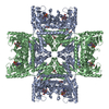 6yb9S S: Starting model for refinement |
|---|---|
| Similar structure data |
-Links
 PDBj
PDBj- Assembly
| Deposited unit | 
| |||||||||||||||||||||
|---|---|---|---|---|---|---|---|---|---|---|---|---|---|---|---|---|---|---|---|---|---|---|
| 1 | 
| |||||||||||||||||||||
| Unit cell |
| |||||||||||||||||||||
| Noncrystallographic symmetry (NCS) | NCS domain:
NCS domain segments: Ens-ID: 1 / Beg auth comp-ID: LEU / Beg label comp-ID: LEU / End auth comp-ID: LEU / End label comp-ID: LEU / Refine code: 0 / Auth seq-ID: 7 - 425 / Label seq-ID: 7 - 425
|
-Components
| #1: Protein | Multi-function printer Mass: 47135.102 Da / Num. of mol.: 2 Source method: isolated from a genetically manipulated source Source: (gene. exp.) Homo sapiens (human) / Gene: PAICS, ADE2, AIRC, PAIS / Production host:  Escherichia coli BL21(DE3) (bacteria) Escherichia coli BL21(DE3) (bacteria)References: UniProt: P22234, phosphoribosylaminoimidazolesuccinocarboxamide synthase, phosphoribosylaminoimidazole carboxylase#2: Chemical |   Mass: 515.424 Da / Num. of mol.: 2 / Source method: obtained synthetically / Formula: C19H27BrN6O4S / Feature type: SUBJECT OF INVESTIGATION Mass: 515.424 Da / Num. of mol.: 2 / Source method: obtained synthetically / Formula: C19H27BrN6O4S / Feature type: SUBJECT OF INVESTIGATION#3: Chemical | Phosphoribosylaminoimidazolesuccinocarboxamide  Mass: 454.283 Da / Num. of mol.: 2 / Source method: obtained synthetically / Formula: C13H19N4O12P / Feature type: SUBJECT OF INVESTIGATION Mass: 454.283 Da / Num. of mol.: 2 / Source method: obtained synthetically / Formula: C13H19N4O12P / Feature type: SUBJECT OF INVESTIGATIONHas ligand of interest | Y | |
|---|
-Experimental details
-Experiment
| Experiment | Method: X-RAY DIFFRACTION / Number of used crystals: 1 |
|---|
- Sample preparation
| Crystal | Density Matthews: 2.74 Å3/Da / Density % sol: 55.05 % / Description: small thin needle |
|---|---|
| Crystal grow | Temperature: 294 K / Method: vapor diffusion, sitting drop / pH: 6.5 Details: 0.1 M MES/imidazole pH 6.5, 10% w/v PEG 8000, 20% v/v ethylene glycol, 0.02 M 1,6-hexanediol, 0.02 M 1-butanol, 0.02 M (RS)-1,2-propanediol, 0.02 M 2-propanol, 0.02 M 1,4-butanediol, and 0.02 M 1,3-propanediol |
-Data collection
| Diffraction | Mean temperature: 100 K / Serial crystal experiment: N | ||||||||||||||||||||||||||||||||||||||||||||||||||||||||||||||||||||||||||||||||||||||||||||||||||||
|---|---|---|---|---|---|---|---|---|---|---|---|---|---|---|---|---|---|---|---|---|---|---|---|---|---|---|---|---|---|---|---|---|---|---|---|---|---|---|---|---|---|---|---|---|---|---|---|---|---|---|---|---|---|---|---|---|---|---|---|---|---|---|---|---|---|---|---|---|---|---|---|---|---|---|---|---|---|---|---|---|---|---|---|---|---|---|---|---|---|---|---|---|---|---|---|---|---|---|---|---|---|
| Diffraction source | Source: SYNCHROTRON / Site: Diamond  / Beamline: I24 / Wavelength: 0.9686 Å / Beamline: I24 / Wavelength: 0.9686 Å | ||||||||||||||||||||||||||||||||||||||||||||||||||||||||||||||||||||||||||||||||||||||||||||||||||||
| Detector | Type: DECTRIS PILATUS3 6M / Detector: PIXEL / Date: May 16, 2019 | ||||||||||||||||||||||||||||||||||||||||||||||||||||||||||||||||||||||||||||||||||||||||||||||||||||
| Radiation | Protocol: SINGLE WAVELENGTH / Monochromatic (M) / Laue (L): M / Scattering type: x-ray | ||||||||||||||||||||||||||||||||||||||||||||||||||||||||||||||||||||||||||||||||||||||||||||||||||||
| Radiation wavelength | Wavelength: 0.9686 Å / Relative weight: 1 | ||||||||||||||||||||||||||||||||||||||||||||||||||||||||||||||||||||||||||||||||||||||||||||||||||||
| Reflection | Resolution: 2.95→49.541 Å / Num. obs: 22274 / % possible obs: 99.9 % / Redundancy: 12.828 % / Biso Wilson estimate: 51.347 Å2 / CC1/2: 0.985 / Rmerge(I) obs: 0.463 / Rrim(I) all: 0.483 / Χ2: 0.637 / Net I/σ(I): 7.01 / Num. measured all: 285722 | ||||||||||||||||||||||||||||||||||||||||||||||||||||||||||||||||||||||||||||||||||||||||||||||||||||
| Reflection shell | Diffraction-ID: 1
|
-Phasing
| Phasing | Method: molecular replacement |
|---|
- Processing
| Software |
| ||||||||||||||||||||||||||||||||||||||||||||||||||||||||||||
|---|---|---|---|---|---|---|---|---|---|---|---|---|---|---|---|---|---|---|---|---|---|---|---|---|---|---|---|---|---|---|---|---|---|---|---|---|---|---|---|---|---|---|---|---|---|---|---|---|---|---|---|---|---|---|---|---|---|---|---|---|---|
| Refinement | Method to determine structure: MOLECULAR REPLACEMENT Starting model: 6yb9 Resolution: 2.95→49.54 Å / Cor.coef. Fo:Fc: 0.908 / Cor.coef. Fo:Fc free: 0.883 / Cross valid method: THROUGHOUT / σ(F): 0 / ESU R Free: 0.439 / Stereochemistry target values: MAXIMUM LIKELIHOOD Details: HYDROGENS HAVE BEEN ADDED IN THE RIDING POSITIONS U VALUES : REFINED INDIVIDUALLY
| ||||||||||||||||||||||||||||||||||||||||||||||||||||||||||||
| Solvent computation | Ion probe radii: 0.8 Å / Shrinkage radii: 0.8 Å / VDW probe radii: 1.2 Å / Solvent model: MASK | ||||||||||||||||||||||||||||||||||||||||||||||||||||||||||||
| Displacement parameters | Biso max: 96.21 Å2 / Biso mean: 23.77 Å2 / Biso min: 3.58 Å2
| ||||||||||||||||||||||||||||||||||||||||||||||||||||||||||||
| Refinement step | Cycle: final / Resolution: 2.95→49.54 Å
| ||||||||||||||||||||||||||||||||||||||||||||||||||||||||||||
| Refine LS restraints |
| ||||||||||||||||||||||||||||||||||||||||||||||||||||||||||||
| Refine LS restraints NCS | Ens-ID: 1 / Number: 12611 / Refine-ID: X-RAY DIFFRACTION / Type: interatomic distance / Rms dev position: 0.12 Å / Weight position: 0.05
| ||||||||||||||||||||||||||||||||||||||||||||||||||||||||||||
| LS refinement shell | Resolution: 2.952→3.028 Å / Rfactor Rfree error: 0 / Total num. of bins used: 20
|
