 Movie
Movie Controller


+ Open data

- Basic information
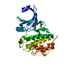
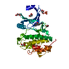
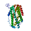
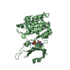
| Entry | Database: PDB / ID: 7akm | ||||||
|---|---|---|---|---|---|---|---|
| Title | Crystal structure of CHK1 kinase domain in complex with ATPyS | ||||||
 Components Components | Serine/threonine-protein kinase Chk1 | ||||||
 Keywords Keywords |  CELL CYCLE CELL CYCLE | ||||||
| Function / homology |  Function and homology information Function and homology informationnegative regulation of G0 to G1 transition / apoptotic process involved in development / histone H3T11 kinase activity / negative regulation of DNA biosynthetic process / negative regulation of mitotic nuclear division / mitotic G2/M transition checkpoint / regulation of mitotic centrosome separation / nucleus organization / inner cell mass cell proliferation / regulation of double-strand break repair via homologous recombination ...negative regulation of G0 to G1 transition / apoptotic process involved in development / histone H3T11 kinase activity / negative regulation of DNA biosynthetic process / negative regulation of mitotic nuclear division / mitotic G2/M transition checkpoint / regulation of mitotic centrosome separation / nucleus organization / inner cell mass cell proliferation / regulation of double-strand break repair via homologous recombination / negative regulation of gene expression, epigenetic / cellular response to caffeine / Transcriptional Regulation by E2F6 / mitotic G2 DNA damage checkpoint signaling / Presynaptic phase of homologous DNA pairing and strand exchange / replicative senescence / Chk1/Chk2(Cds1) mediated inactivation of Cyclin B:Cdk1 complex / positive regulation of cell cycle / Activation of ATR in response to replication stress / signal transduction in response to DNA damage / DNA damage checkpoint signaling / replication fork / regulation of signal transduction by p53 class mediator / condensed nuclear chromosome / Ubiquitin Mediated Degradation of Phosphorylated Cdc25A / TP53 Regulates Transcription of DNA Repair Genes / peptidyl-threonine phosphorylation / G2/M DNA damage checkpoint / Signaling by SCF-KIT / cellular response to mechanical stimulus / G2/M transition of mitotic cell cycle / regulation of cell population proliferation / Processing of DNA double-strand break ends / Regulation of TP53 Activity through Phosphorylation / DNA replication / non-specific serine/threonine protein kinase / protein kinase activity / intracellular signal transduction / chromatin remodeling / protein domain specific binding / protein phosphorylation / protein serine kinase activity / intracellular membrane-bounded organelle / DNA repair / protein serine/threonine kinase activity / centrosome / apoptotic process / DNA damage response / chromatin / protein-containing complex / extracellular space / nucleoplasm / ATP binding / nucleus / cytosol / cytoplasmSimilarity search - Function | ||||||
| Biological species |  Homo sapiens (human) Homo sapiens (human) | ||||||
| Method | X-RAY DIFFRACTION / SYNCHROTRON / MOLECULAR REPLACEMENT / Resolution: 1.93 Å | ||||||
 Authors Authors | Day, M. / Oliver, A.W. / Pearl, L.H. | ||||||
| Funding support |  United Kingdom, 1items United Kingdom, 1items
| ||||||
 Citation Citation | Journal: Structure / Year: 2021 Title: Structural basis for recruitment of the CHK1 DNA damage kinase by the CLASPIN scaffold protein. Authors: Day, M. / Parry-Morris, S. / Houghton-Gisby, J. / Oliver, A.W. / Pearl, L.H. | ||||||
| History |
|
- Structure visualization
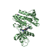
| Structure viewer | Molecule: MolmilJmol/JSmol |
|---|
- Downloads & links
-Download
| PDBx/mmCIF format | 7akm.cif.gz | 284.3 KB | Display | PDBx/mmCIF format |
|---|---|---|---|---|
| PDB format | pdb7akm.ent.gz | 184.9 KB | Display | PDB format |
| PDBx/mmJSON format | 7akm.json.gz | Tree view | PDBx/mmJSON format | |
| Others |  Other downloads Other downloads |
-Validation report
| Arichive directory | https://data.pdbj.org/pub/pdb/validation_reports/ak/7akmftp://data.pdbj.org/pub/pdb/validation_reports/ak/7akm | HTTPS FTP |
|---|
-Related structure data
| Related structure data |  7akoC 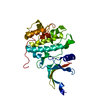 1ia8S S: Starting model for refinement C: citing same article ( |
|---|---|
| Similar structure data |


-Links
 PDBj
PDBj


- Assembly
| Deposited unit | 
| ||||||||||||
|---|---|---|---|---|---|---|---|---|---|---|---|---|---|
| 1 | 
| ||||||||||||
| 2 | 
| ||||||||||||
| Unit cell |
|
-Components
-Protein , 1 types, 2 molecules AB
| #1: Protein | Mass: 34213.219 Da / Num. of mol.: 2 / Mutation: D10R Source method: isolated from a genetically manipulated source Source: (gene. exp.) Homo sapiens (human) / Gene: CHEK1, CHK1 / Plasmid: pFASTBAC / Production host:   Spodoptera frugiperda (fall armyworm) Spodoptera frugiperda (fall armyworm)References: UniProt: O14757, non-specific serine/threonine protein kinase |
|---|
-Non-polymers , 5 types, 448 molecules 


| #2: Chemical | ChemComp-AGS /  Mass: 523.247 Da / Num. of mol.: 1 / Source method: obtained synthetically / Formula: C10H16N5O12P3S / Feature type: SUBJECT OF INVESTIGATION / Comment: ATP-gamma-S, energy-carrying molecule analogue*YM Mass: 523.247 Da / Num. of mol.: 1 / Source method: obtained synthetically / Formula: C10H16N5O12P3S / Feature type: SUBJECT OF INVESTIGATION / Comment: ATP-gamma-S, energy-carrying molecule analogue*YM | ||||||
|---|---|---|---|---|---|---|---|
| #3: Chemical |  Mass: 24.305 Da / Num. of mol.: 3 / Source method: obtained synthetically / Formula: Mg Mass: 24.305 Da / Num. of mol.: 3 / Source method: obtained synthetically / Formula: Mg#4: Chemical | ChemComp-EDO / Ethylene glycol Mass: 62.068 Da / Num. of mol.: 9 / Source method: obtained synthetically / Formula: C2H6O2 Mass: 62.068 Da / Num. of mol.: 9 / Source method: obtained synthetically / Formula: C2H6O2#5: Chemical | ChemComp-CIT / | Citric acid Mass: 192.124 Da / Num. of mol.: 1 / Source method: isolated from a natural source / Formula: C6H8O7 Mass: 192.124 Da / Num. of mol.: 1 / Source method: isolated from a natural source / Formula: C6H8O7#6: Water | ChemComp-HOH / | WaterMass: 18.015 Da / Num. of mol.: 434 / Source method: isolated from a natural source / Formula: H2O |
-Details
| Has ligand of interest | Y |
|---|
-Experimental details
-Experiment
| Experiment | Method: X-RAY DIFFRACTION / Number of used crystals: 1 |
|---|
- Sample preparation
| Crystal | Density Matthews: 2.62 Å3/Da / Density % sol: 52.97 % |
|---|---|
| Crystal grow | Temperature: 287.15 K / Method: vapor diffusion, sitting drop / pH: 8.5 Details: 200 mM Sodium citrate tribasic dihydrate, 100 mM Bis-Tris propane pH 8.5 and 20% w/v PEG 3350 |
-Data collection
| Diffraction | Mean temperature: 100 K / Serial crystal experiment: N |
|---|---|
| Diffraction source | Source: SYNCHROTRON / Site: Diamond / Beamline: I24 / Wavelength: 0.96864 Å |
| Detector | Type: DECTRIS PILATUS3 6M / Detector: PIXEL / Date: Feb 19, 2020 |
| Radiation | Protocol: SINGLE WAVELENGTH / Monochromatic (M) / Laue (L): M / Scattering type: x-ray |
| Radiation wavelength | Wavelength: 0.96864 Å / Relative weight: 1 |
| Reflection | Resolution: 1.93→44.63 Å / Num. obs: 49422 / % possible obs: 98.35 % / Redundancy: 4 % / Biso Wilson estimate: 28.6 Å2 / CC1/2: 0.992 / Rmerge(I) obs: 0.1555 / Rpim(I) all: 0.08786 / Rrim(I) all: 0.1795 / Net I/σ(I): 5.57 |
| Reflection shell | Resolution: 1.93→1.999 Å / Rmerge(I) obs: 1.313 / Mean I/σ(I) obs: 0.79 / Num. unique obs: 4447 / CC1/2: 0.336 / Rpim(I) all: 0.7813 / Rsym value: 1.54 |
- Processing
| Software |
| ||||||||||||||||||||||||||||||||||||||||||||||||||||||||||||||||||||||||||||||||||||||||||||||||||||||||||||||||||||||||||||||
|---|---|---|---|---|---|---|---|---|---|---|---|---|---|---|---|---|---|---|---|---|---|---|---|---|---|---|---|---|---|---|---|---|---|---|---|---|---|---|---|---|---|---|---|---|---|---|---|---|---|---|---|---|---|---|---|---|---|---|---|---|---|---|---|---|---|---|---|---|---|---|---|---|---|---|---|---|---|---|---|---|---|---|---|---|---|---|---|---|---|---|---|---|---|---|---|---|---|---|---|---|---|---|---|---|---|---|---|---|---|---|---|---|---|---|---|---|---|---|---|---|---|---|---|---|---|---|---|
| Refinement | Method to determine structure: MOLECULAR REPLACEMENT Starting model: 1IA8 Resolution: 1.93→44.63 Å / SU ML: 0.2459 / Cross valid method: FREE R-VALUE / σ(F): 1.34 / Phase error: 25.2472
| ||||||||||||||||||||||||||||||||||||||||||||||||||||||||||||||||||||||||||||||||||||||||||||||||||||||||||||||||||||||||||||||
| Solvent computation | Shrinkage radii: 0.9 Å / VDW probe radii: 1.11 Å | ||||||||||||||||||||||||||||||||||||||||||||||||||||||||||||||||||||||||||||||||||||||||||||||||||||||||||||||||||||||||||||||
| Displacement parameters | Biso mean: 36.48 Å2 | ||||||||||||||||||||||||||||||||||||||||||||||||||||||||||||||||||||||||||||||||||||||||||||||||||||||||||||||||||||||||||||||
| Refinement step | Cycle: LAST / Resolution: 1.93→44.63 Å
| ||||||||||||||||||||||||||||||||||||||||||||||||||||||||||||||||||||||||||||||||||||||||||||||||||||||||||||||||||||||||||||||
| Refine LS restraints |
| ||||||||||||||||||||||||||||||||||||||||||||||||||||||||||||||||||||||||||||||||||||||||||||||||||||||||||||||||||||||||||||||
| LS refinement shell |
|
