 Movie
Movie Controller


+ Open data

- Basic information
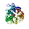
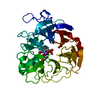
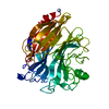
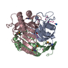
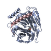
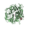
| Entry | Database: PDB / ID: 7af2 | ||||||
|---|---|---|---|---|---|---|---|
| Title | Salmonella typhimurium neuraminidase mutant (D62G) | ||||||
 Components Components | Sialidase Neuraminidase Neuraminidase | ||||||
 Keywords Keywords | HYDROLASE / Salmonella typhimurium / enzyme / neuraminidase / mutant / D62G / sialidase / native / hydrolase. | ||||||
| Function / homology |  Function and homology informationexo-alpha-sialidase activity / ganglioside catabolic process / oligosaccharide catabolic process / exo-alpha-(2->3)-sialidase activity / exo-alpha-(2->6)-sialidase activity / exo-alpha-(2->8)-sialidase activity / exo-alpha-sialidase / intracellular membrane-bounded organelle / membrane / cytoplasm Function and homology informationexo-alpha-sialidase activity / ganglioside catabolic process / oligosaccharide catabolic process / exo-alpha-(2->3)-sialidase activity / exo-alpha-(2->6)-sialidase activity / exo-alpha-(2->8)-sialidase activity / exo-alpha-sialidase / intracellular membrane-bounded organelle / membrane / cytoplasmSimilarity search - Function | ||||||
| Biological species |  Salmonella enterica subsp. enterica serovar Typhimurium (bacteria) Salmonella enterica subsp. enterica serovar Typhimurium (bacteria) | ||||||
| Method | X-RAY DIFFRACTION / SYNCHROTRON / MOLECULAR REPLACEMENT / Resolution: 0.792 Å | ||||||
 Authors Authors | Salinger, M.T. / Kuhn, P. / Laver, W.G. / Pape, T. / Schneider, T.R. / Sheldrick, G.M. / Vimr, E.R. / Garman, E.F. | ||||||
 Citation Citation | Journal: To Be Published Title: Salmonella typhimurium neuraminidase mutant (D62G) Authors: Garman, E.F. / Salinger, M.T. / Laver, W.G. / Kuhn, P. / Pape, T. / Schneider, T.R. / Sheldrick, G.M. / Vimr, E.R. | ||||||
| History |
|
- Structure visualization
| Structure viewer | Molecule: MolmilJmol/JSmol |
|---|
- Downloads & links
-Download
| PDBx/mmCIF format | 7af2.cif.gz | 286.1 KB | Display | PDBx/mmCIF format |
|---|---|---|---|---|
| PDB format | pdb7af2.ent.gz | Display | PDB format | |
| PDBx/mmJSON format | 7af2.json.gz | Tree view | PDBx/mmJSON format | |
| Others |  Other downloads Other downloads |
-Validation report
| Arichive directory | https://data.pdbj.org/pub/pdb/validation_reports/af/7af2ftp://data.pdbj.org/pub/pdb/validation_reports/af/7af2 | HTTPS FTP |
|---|
-Related structure data
| Related structure data |  3silS S: Starting model for refinement |
|---|---|
| Similar structure data |


-Links
 PDBj
PDBj

- Assembly
| Deposited unit | 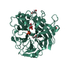
| ||||||||
|---|---|---|---|---|---|---|---|---|---|
| 1 |
| ||||||||
| Unit cell |
|
-Components
| #1: Protein | Neuraminidase / N-acylneuraminate glycohydrolase / Neuraminidase / NANase / STNA Mass: 41733.520 Da / Num. of mol.: 1 / Mutation: D62G Source method: isolated from a genetically manipulated source Source: (gene. exp.) Salmonella enterica subsp. enterica serovar Typhimurium (bacteria)Gene: nanH, STM0928 / Production host: Escherichia coli (E. coli) / References: UniProt: P29768, exo-alpha-sialidase | ||||||
|---|---|---|---|---|---|---|---|
| #2: Chemical | ChemComp-GOL / Glycerol  Mass: 92.094 Da / Num. of mol.: 5 / Source method: obtained synthetically / Formula: C3H8O3 Mass: 92.094 Da / Num. of mol.: 5 / Source method: obtained synthetically / Formula: C3H8O3#3: Chemical | ChemComp-PO4 / | Phosphate  Mass: 94.971 Da / Num. of mol.: 1 / Source method: obtained synthetically / Formula: PO4 Mass: 94.971 Da / Num. of mol.: 1 / Source method: obtained synthetically / Formula: PO4#4: Water | ChemComp-HOH / | Water Mass: 18.015 Da / Num. of mol.: 502 / Source method: isolated from a natural source / Formula: H2O Mass: 18.015 Da / Num. of mol.: 502 / Source method: isolated from a natural source / Formula: H2OHas ligand of interest | N | |
-Experimental details
-Experiment
| Experiment | Method: X-RAY DIFFRACTION / Number of used crystals: 1 |
|---|
- Sample preparation
| Crystal | Density Matthews: 2.08 Å3/Da / Density % sol: 40.92 % |
|---|---|
| Crystal grow | Temperature: 277 K / Method: vapor diffusion, hanging drop Details: Crystals grown by hanging drop vapour diffusion. A 1:1 mixture of protein solution and an 8:4 mixture of K2HPO4 to KH2PO4 was placed above a well of an 8:6 solution of K2HPO4 to KH2PO4. Then ...Details: Crystals grown by hanging drop vapour diffusion. A 1:1 mixture of protein solution and an 8:4 mixture of K2HPO4 to KH2PO4 was placed above a well of an 8:6 solution of K2HPO4 to KH2PO4. Then serially cryoprotected in situ to 40% glycerol (v/v with mother liquor) in 10% increments over a period of a few minutes. |
-Data collection
| Diffraction | Mean temperature: 100 K / Serial crystal experiment: N |
|---|---|
| Diffraction source | Source: SYNCHROTRON / Site: SSRL  / Beamline: BL9-1 / Wavelength: 0.79 Å / Beamline: BL9-1 / Wavelength: 0.79 Å |
| Detector | Type: MAR scanner 345 mm plate / Detector: IMAGE PLATE / Date: Feb 27, 1998 |
| Radiation | Protocol: SINGLE WAVELENGTH / Monochromatic (M) / Laue (L): M / Scattering type: x-ray |
| Radiation wavelength | Wavelength: 0.79 Å / Relative weight: 1 |
| Reflection | Resolution: 0.792→18.107 Å / Num. obs: 355798 / % possible obs: 94.7 % / Redundancy: 3.2 % / Rrim(I) all: 0.061 / Rsym value: 0.053 / Net I/σ(I): 23.61 |
| Reflection shell | Resolution: 0.792→0.81 Å / Redundancy: 1.8 % / Mean I/σ(I) obs: 0.88 / Num. unique obs: 19346 / Rrim(I) all: 1.572 / Rsym value: 1.161 / % possible all: 70.1 |
- Processing
| Software |
| ||||||||||||||||||||||||||||||||||||||||||||||||||||||||||||||||||||||||||||||||||||||||||||||||||||||||||||||||||||||||||||||||||||||||||||||||||||||||||||||||
|---|---|---|---|---|---|---|---|---|---|---|---|---|---|---|---|---|---|---|---|---|---|---|---|---|---|---|---|---|---|---|---|---|---|---|---|---|---|---|---|---|---|---|---|---|---|---|---|---|---|---|---|---|---|---|---|---|---|---|---|---|---|---|---|---|---|---|---|---|---|---|---|---|---|---|---|---|---|---|---|---|---|---|---|---|---|---|---|---|---|---|---|---|---|---|---|---|---|---|---|---|---|---|---|---|---|---|---|---|---|---|---|---|---|---|---|---|---|---|---|---|---|---|---|---|---|---|---|---|---|---|---|---|---|---|---|---|---|---|---|---|---|---|---|---|---|---|---|---|---|---|---|---|---|---|---|---|---|---|---|---|---|
| Refinement | Method to determine structure: MOLECULAR REPLACEMENT Starting model: 3SIL Resolution: 0.792→18.102 Å / Cor.coef. Fo:Fc: 0.987 / Cor.coef. Fo:Fc free: 0.986 / WRfactor Rfree: 0.14 / WRfactor Rwork: 0.126 / Average fsc free: 0.8265 / Average fsc work: 0.829 / Cross valid method: FREE R-VALUE / ESU R: 0.011 / ESU R Free: 0.012 Details: Hydrogens have been added in their riding positions
| ||||||||||||||||||||||||||||||||||||||||||||||||||||||||||||||||||||||||||||||||||||||||||||||||||||||||||||||||||||||||||||||||||||||||||||||||||||||||||||||||
| Solvent computation | Ion probe radii: 0.8 Å / Shrinkage radii: 0.8 Å / VDW probe radii: 1.2 Å / Solvent model: MASK BULK SOLVENT | ||||||||||||||||||||||||||||||||||||||||||||||||||||||||||||||||||||||||||||||||||||||||||||||||||||||||||||||||||||||||||||||||||||||||||||||||||||||||||||||||
| Displacement parameters | Biso mean: 15.115 Å2
| ||||||||||||||||||||||||||||||||||||||||||||||||||||||||||||||||||||||||||||||||||||||||||||||||||||||||||||||||||||||||||||||||||||||||||||||||||||||||||||||||
| Refinement step | Cycle: LAST / Resolution: 0.792→18.102 Å
| ||||||||||||||||||||||||||||||||||||||||||||||||||||||||||||||||||||||||||||||||||||||||||||||||||||||||||||||||||||||||||||||||||||||||||||||||||||||||||||||||
| Refine LS restraints |
| ||||||||||||||||||||||||||||||||||||||||||||||||||||||||||||||||||||||||||||||||||||||||||||||||||||||||||||||||||||||||||||||||||||||||||||||||||||||||||||||||
| LS refinement shell |
|
