 Movie
Movie Controller

[English] 日本語
 Yorodumi
- PDB-7aau: Crystal structure of nitrosoglutathione reductase from Chlamydomo... -
+ Open data

- Basic information
| Entry | Database: PDB / ID: 7aau | |||||||||
|---|---|---|---|---|---|---|---|---|---|---|
| Title | Crystal structure of nitrosoglutathione reductase from Chlamydomonas reinhardtii in complex with NAD+ | |||||||||
 Components Components | S-(hydroxymethyl)glutathione dehydrogenase S-(hydroxymethyl)glutathione dehydrogenase S-(hydroxymethyl)glutathione dehydrogenase | |||||||||
 Keywords Keywords | OXIDOREDUCTASE / Alcohol dehydrogenase / Zinc-binding dehydrogenase | |||||||||
| Function / homology |  Function and homology informationS-(hydroxymethyl)glutathione dehydrogenase / S-(hydroxymethyl)glutathione dehydrogenase (NADP+) activity / S-(hydroxymethyl)glutathione dehydrogenase (NAD+) activity / S-(hydroxymethyl)glutathione dehydrogenase (NAD(P)+) activity / formaldehyde catabolic process / alcohol dehydrogenase (NAD+) activity, zinc-dependent / : / zinc ion binding / cytosol Function and homology informationS-(hydroxymethyl)glutathione dehydrogenase / S-(hydroxymethyl)glutathione dehydrogenase (NADP+) activity / S-(hydroxymethyl)glutathione dehydrogenase (NAD+) activity / S-(hydroxymethyl)glutathione dehydrogenase (NAD(P)+) activity / formaldehyde catabolic process / alcohol dehydrogenase (NAD+) activity, zinc-dependent / : / zinc ion binding / cytosolSimilarity search - Function | |||||||||
| Biological species |   Chlamydomonas reinhardtii (plant) Chlamydomonas reinhardtii (plant) | |||||||||
| Method | X-RAY DIFFRACTION / SYNCHROTRON / MOLECULAR REPLACEMENT / Resolution: 2.301 Å | |||||||||
 Authors Authors | Fermani, S. / Zaffagnini, M. / Falini, G. / Lemaire, S.D. | |||||||||
| Funding support |  Italy, Italy,  France, 2items France, 2items
| |||||||||
 Citation Citation | Journal: Redox Biol / Year: 2020 Title: Structural and functional insights into nitrosoglutathione reductase from Chlamydomonas reinhardtii. Authors: Tagliani, A. / Rossi, J. / Marchand, C.H. / De Mia, M. / Tedesco, D. / Gurrieri, L. / Meloni, M. / Falini, G. / Trost, P. / Lemaire, S.D. / Fermani, S. / Zaffagnini, M. | |||||||||
| History |
|
- Structure visualization
| Structure viewer | Molecule: MolmilJmol/JSmol |
|---|
- Downloads & links
-Download
| PDBx/mmCIF format | 7aau.cif.gz | 461.8 KB | Display | PDBx/mmCIF format |
|---|---|---|---|---|
| PDB format | pdb7aau.ent.gz | 378.9 KB | Display | PDB format |
| PDBx/mmJSON format | 7aau.json.gz | Tree view | PDBx/mmJSON format | |
| Others |  Other downloads Other downloads |
-Validation report
| Arichive directory | https://data.pdbj.org/pub/pdb/validation_reports/aa/7aauftp://data.pdbj.org/pub/pdb/validation_reports/aa/7aau | HTTPS FTP |
|---|
-Related structure data
| Related structure data |  7aasC 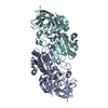 7av7C  4dlaS S: Starting model for refinement C: citing same article ( |
|---|---|
| Similar structure data |


-Links
 PDBj
PDBj

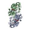
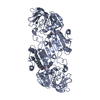
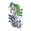
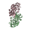
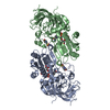
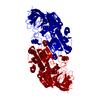
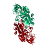
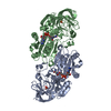
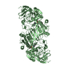
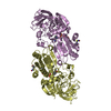
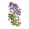
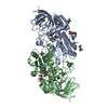
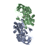
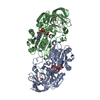
- Assembly
| Deposited unit | 
| ||||||||
|---|---|---|---|---|---|---|---|---|---|
| 1 | 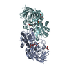
| ||||||||
| 2 | 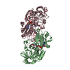
| ||||||||
| 3 | 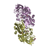
| ||||||||
| Unit cell |
|
-Components
-Protein , 1 types, 6 molecules ABCDEF
| #1: Protein | S-(hydroxymethyl)glutathione dehydrogenase Mass: 40455.410 Da / Num. of mol.: 6 Source method: isolated from a genetically manipulated source Source: (gene. exp.) Chlamydomonas reinhardtii (plant) / Gene: CHLRE_12g543400v5 / Plasmid: pET3C / Production host:  Escherichia coli (E. coli) Escherichia coli (E. coli)References: UniProt: A0A2K3D6R4, S-(hydroxymethyl)glutathione dehydrogenase |
|---|
-Non-polymers , 6 types, 1491 molecules 


| #2: Chemical | ChemComp-ZN /  Mass: 65.409 Da / Num. of mol.: 12 / Source method: obtained synthetically / Formula: Zn Mass: 65.409 Da / Num. of mol.: 12 / Source method: obtained synthetically / Formula: Zn#3: Chemical | ChemComp-NAD / Nicotinamide adenine dinucleotide Mass: 663.425 Da / Num. of mol.: 6 / Source method: obtained synthetically / Formula: C21H27N7O14P2 / Feature type: SUBJECT OF INVESTIGATION / Comment: NAD*YM Mass: 663.425 Da / Num. of mol.: 6 / Source method: obtained synthetically / Formula: C21H27N7O14P2 / Feature type: SUBJECT OF INVESTIGATION / Comment: NAD*YM#4: Chemical | ChemComp-PEG / Diethylene glycol Mass: 106.120 Da / Num. of mol.: 15 / Source method: obtained synthetically / Formula: C4H10O3 Mass: 106.120 Da / Num. of mol.: 15 / Source method: obtained synthetically / Formula: C4H10O3#5: Chemical | ChemComp-CL / Chloride Mass: 35.453 Da / Num. of mol.: 4 / Source method: obtained synthetically / Formula: Cl Mass: 35.453 Da / Num. of mol.: 4 / Source method: obtained synthetically / Formula: Cl#6: Chemical | ChemComp-MG / |  Mass: 24.305 Da / Num. of mol.: 1 / Source method: obtained synthetically / Formula: Mg Mass: 24.305 Da / Num. of mol.: 1 / Source method: obtained synthetically / Formula: Mg#7: Water | ChemComp-HOH / | WaterMass: 18.015 Da / Num. of mol.: 1453 / Source method: isolated from a natural source / Formula: H2O |
|---|
-Details
| Has ligand of interest | Y |
|---|
-Experimental details
-Experiment
| Experiment | Method: X-RAY DIFFRACTION / Number of used crystals: 1 |
|---|
- Sample preparation
| Crystal | Density Matthews: 2.4 Å3/Da / Density % sol: 48 % / Description: stick crystals |
|---|---|
| Crystal grow | Temperature: 293 K / Method: vapor diffusion, hanging drop / pH: 8.5 Details: 0.1 M Tris-HCl pH 8.5, 0.1 M MgCl2 or Mg(CH3CO2)2, and 12-15% w/v PEG 20K or 12% w/v PEG 8K |
-Data collection
| Diffraction | Mean temperature: 100 K / Serial crystal experiment: N | |||||||||||||||||||||||||||
|---|---|---|---|---|---|---|---|---|---|---|---|---|---|---|---|---|---|---|---|---|---|---|---|---|---|---|---|---|
| Diffraction source | Source: SYNCHROTRON / Site: ELETTRA / Beamline: 5.2R / Wavelength: 0.9137 Å | |||||||||||||||||||||||||||
| Detector | Type: DECTRIS PILATUS 2M / Detector: PIXEL / Date: Aug 4, 2015 | |||||||||||||||||||||||||||
| Radiation | Protocol: SINGLE WAVELENGTH / Monochromatic (M) / Laue (L): M / Scattering type: x-ray | |||||||||||||||||||||||||||
| Radiation wavelength | Wavelength: 0.9137 Å / Relative weight: 1 | |||||||||||||||||||||||||||
| Reflection | Resolution: 2.3→117.4 Å / Num. obs: 102418 / % possible obs: 99.7 % / Redundancy: 4.2 % / Biso Wilson estimate: 22.16 Å2 / CC1/2: 0.988 / Rmerge(I) obs: 0.157 / Rpim(I) all: 0.087 / Rrim(I) all: 0.18 / Net I/σ(I): 8.3 | |||||||||||||||||||||||||||
| Reflection shell | Diffraction-ID: 1
|
- Processing
| Software |
| ||||||||||||||||||||||||||||||||||||||||||||||||||||||||||||||||||||||||||||||||||||||||||||||||||||||||||||||||||||||||||||||||||||||||||||||||||||||||||||||||||||||||||||||||||||||||||
|---|---|---|---|---|---|---|---|---|---|---|---|---|---|---|---|---|---|---|---|---|---|---|---|---|---|---|---|---|---|---|---|---|---|---|---|---|---|---|---|---|---|---|---|---|---|---|---|---|---|---|---|---|---|---|---|---|---|---|---|---|---|---|---|---|---|---|---|---|---|---|---|---|---|---|---|---|---|---|---|---|---|---|---|---|---|---|---|---|---|---|---|---|---|---|---|---|---|---|---|---|---|---|---|---|---|---|---|---|---|---|---|---|---|---|---|---|---|---|---|---|---|---|---|---|---|---|---|---|---|---|---|---|---|---|---|---|---|---|---|---|---|---|---|---|---|---|---|---|---|---|---|---|---|---|---|---|---|---|---|---|---|---|---|---|---|---|---|---|---|---|---|---|---|---|---|---|---|---|---|---|---|---|---|---|---|---|---|
| Refinement | Method to determine structure: MOLECULAR REPLACEMENT Starting model: 4DLA Resolution: 2.301→48.565 Å / SU ML: 0.27 / Cross valid method: FREE R-VALUE / σ(F): 0.13 / Phase error: 23.33 / Stereochemistry target values: ML
| ||||||||||||||||||||||||||||||||||||||||||||||||||||||||||||||||||||||||||||||||||||||||||||||||||||||||||||||||||||||||||||||||||||||||||||||||||||||||||||||||||||||||||||||||||||||||||
| Solvent computation | Shrinkage radii: 0.9 Å / VDW probe radii: 1.11 Å / Solvent model: FLAT BULK SOLVENT MODEL | ||||||||||||||||||||||||||||||||||||||||||||||||||||||||||||||||||||||||||||||||||||||||||||||||||||||||||||||||||||||||||||||||||||||||||||||||||||||||||||||||||||||||||||||||||||||||||
| Displacement parameters | Biso max: 91.48 Å2 / Biso mean: 31.6457 Å2 / Biso min: 12.89 Å2 | ||||||||||||||||||||||||||||||||||||||||||||||||||||||||||||||||||||||||||||||||||||||||||||||||||||||||||||||||||||||||||||||||||||||||||||||||||||||||||||||||||||||||||||||||||||||||||
| Refinement step | Cycle: final / Resolution: 2.301→48.565 Å
| ||||||||||||||||||||||||||||||||||||||||||||||||||||||||||||||||||||||||||||||||||||||||||||||||||||||||||||||||||||||||||||||||||||||||||||||||||||||||||||||||||||||||||||||||||||||||||
| LS refinement shell | Refine-ID: X-RAY DIFFRACTION / Rfactor Rfree error: 0
|
