 Movie
Movie Controller

[English] 日本語
 Yorodumi
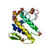
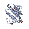
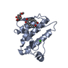
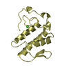
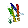
- PDB-6ztk: Crystal structure of Mialostatin, a gut cystatin from the hard ti... -
+ Open data

- Basic information
| Entry | Database: PDB / ID: 6ztk | |||||||||
|---|---|---|---|---|---|---|---|---|---|---|
| Title | Crystal structure of Mialostatin, a gut cystatin from the hard tick Ixodes ricinus | |||||||||
 Components Components | Mialostatin | |||||||||
 Keywords Keywords |  PROTEIN BINDING / cystatin / protease inhibitor / ixodes ricinus / tick / hydrolase inhibitor PROTEIN BINDING / cystatin / protease inhibitor / ixodes ricinus / tick / hydrolase inhibitor | |||||||||
| Function / homology | Cystatin domain / Cystatin-like domain / Cystatin domain / cysteine-type endopeptidase inhibitor activity / Putative tick cistatins 1 Function and homology information Function and homology information | |||||||||
| Biological species |  Ixodes ricinus (castor bean tick) Ixodes ricinus (castor bean tick) | |||||||||
| Method | X-RAY DIFFRACTION / SYNCHROTRON / MOLECULAR REPLACEMENT / Resolution: 1.55 Å | |||||||||
 Authors Authors | Busa, M. / Rezacova, P. / Mares, M. | |||||||||
| Funding support | European Union,  Czech Republic, 2items Czech Republic, 2items
| |||||||||
 Citation Citation | Journal: Int J Mol Sci / Year: 2021 Title: Mialostatin, a Novel Midgut Cystatin from Ixodes ricinus Ticks: Crystal Structure and Regulation of Host Blood Digestion. Authors: Kotal, J. / Busa, M. / Urbanova, V. / Rezacova, P. / Chmelar, J. / Langhansova, H. / Sojka, D. / Mares, M. / Kotsyfakis, M. | |||||||||
| History |
|
- Structure visualization
| Structure viewer | Molecule: MolmilJmol/JSmol |
|---|
- Downloads & links
-Download
| PDBx/mmCIF format | 6ztk.cif.gz | 71.6 KB | Display | PDBx/mmCIF format |
|---|---|---|---|---|
| PDB format | pdb6ztk.ent.gz | 51.1 KB | Display | PDB format |
| PDBx/mmJSON format | 6ztk.json.gz | Tree view | PDBx/mmJSON format | |
| Others |  Other downloads Other downloads |
-Validation report
| Arichive directory | https://data.pdbj.org/pub/pdb/validation_reports/zt/6ztkftp://data.pdbj.org/pub/pdb/validation_reports/zt/6ztk | HTTPS FTP |
|---|
-Related structure data
| Related structure data | 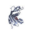 3l0rS S: Starting model for refinement |
|---|---|
| Similar structure data |


-Links
 PDBj
PDBj
- Assembly
| Deposited unit | 
| ||||||||
|---|---|---|---|---|---|---|---|---|---|
| 1 | 
| ||||||||
| 2 | 
| ||||||||
| Unit cell |
| ||||||||
| Components on special symmetry positions |
|
-Components
| #1: Protein | Mass: 15768.985 Da / Num. of mol.: 2 Source method: isolated from a genetically manipulated source Details: Mialostatin / Source: (gene. exp.) Ixodes ricinus (castor bean tick) / Production host:  Escherichia coli (E. coli) / References: UniProt: V5H924 Escherichia coli (E. coli) / References: UniProt: V5H924#2: Chemical | Triton X-100  Mass: 352.508 Da / Num. of mol.: 2 / Source method: obtained synthetically / Formula: C21H36O4 / Comment: detergent*YM Mass: 352.508 Da / Num. of mol.: 2 / Source method: obtained synthetically / Formula: C21H36O4 / Comment: detergent*YM#3: Chemical | Sulfate  Mass: 96.063 Da / Num. of mol.: 3 / Source method: obtained synthetically / Formula: SO4 Mass: 96.063 Da / Num. of mol.: 3 / Source method: obtained synthetically / Formula: SO4#4: Water | ChemComp-HOH / | Water Mass: 18.015 Da / Num. of mol.: 369 / Source method: isolated from a natural source / Formula: H2O Mass: 18.015 Da / Num. of mol.: 369 / Source method: isolated from a natural source / Formula: H2OHas ligand of interest | N | |
|---|
-Experimental details
-Experiment
| Experiment | Method: X-RAY DIFFRACTION / Number of used crystals: 1 |
|---|
- Sample preparation
| Crystal | Density Matthews: 2.44 Å3/Da / Density % sol: 49.61 % / Description: hexagonal prism |
|---|---|
| Crystal grow | Temperature: 291.15 K / Method: vapor diffusion, hanging drop / pH: 4 / Details: 1M ammonium sulphate 0.1M sodium citrate pH 4.0 |
-Data collection
| Diffraction | Mean temperature: 100 K / Serial crystal experiment: N |
|---|---|
| Diffraction source | Source: SYNCHROTRON / Site: BESSY  / Beamline: 14.1 / Wavelength: 0.91841 Å / Beamline: 14.1 / Wavelength: 0.91841 Å |
| Detector | Type: DECTRIS PILATUS 6M / Detector: PIXEL / Date: Mar 17, 2016 |
| Radiation | Protocol: SINGLE WAVELENGTH / Monochromatic (M) / Laue (L): M / Scattering type: x-ray |
| Radiation wavelength | Wavelength: 0.91841 Å / Relative weight: 1 |
| Reflection | Resolution: 1.55→42.9 Å / Num. obs: 46381 / % possible obs: 99.9 % / Redundancy: 21.22 % / Biso Wilson estimate: 19.54 Å2 / CC1/2: 1 / CC star: 1 / Rmerge(I) obs: 0.08446 / Rpim(I) all: 0.01874 / Rrim(I) all: 0.087 / Net I/av σ(I): 17.13 / Net I/σ(I): 21.95 |
| Reflection shell | Resolution: 1.55→1.65 Å / Redundancy: 21.55 % / Rmerge(I) obs: 1.203 / Mean I/σ(I) obs: 2 / Num. unique obs: 4528 / CC1/2: 0.764 / CC star: 0.936 / Rpim(I) all: 0.263 / Rrim(I) all: 1.232 / % possible all: 99.25 |
- Processing
| Software |
| ||||||||||||||||||||||||||||||||||||||||||||||||||||||||||||
|---|---|---|---|---|---|---|---|---|---|---|---|---|---|---|---|---|---|---|---|---|---|---|---|---|---|---|---|---|---|---|---|---|---|---|---|---|---|---|---|---|---|---|---|---|---|---|---|---|---|---|---|---|---|---|---|---|---|---|---|---|---|
| Refinement | Method to determine structure: MOLECULAR REPLACEMENT Starting model: 3L0R Resolution: 1.55→42.9 Å / Cor.coef. Fo:Fc: 0.969 / Cor.coef. Fo:Fc free: 0.958 / SU B: 1.266 / SU ML: 0.046 / Cross valid method: THROUGHOUT / σ(F): 0 / ESU R: 0.068 / ESU R Free: 0.072 Details: HYDROGENS HAVE BEEN ADDED IN THE RIDING POSITIONS U VALUES : REFINED INDIVIDUALLY
| ||||||||||||||||||||||||||||||||||||||||||||||||||||||||||||
| Solvent computation | Ion probe radii: 0.8 Å / Shrinkage radii: 0.8 Å / VDW probe radii: 1.2 Å | ||||||||||||||||||||||||||||||||||||||||||||||||||||||||||||
| Displacement parameters | Biso max: 108.8 Å2 / Biso mean: 20.97 Å2 / Biso min: 11.36 Å2
| ||||||||||||||||||||||||||||||||||||||||||||||||||||||||||||
| Refinement step | Cycle: final / Resolution: 1.55→42.9 Å
| ||||||||||||||||||||||||||||||||||||||||||||||||||||||||||||
| Refine LS restraints |
| ||||||||||||||||||||||||||||||||||||||||||||||||||||||||||||
| LS refinement shell | Resolution: 1.552→1.592 Å / Rfactor Rfree error: 0 / Total num. of bins used: 20
|
