 Movie
Movie Controller

[English] 日本語
 Yorodumi
- PDB-6zje: Crystal structure of human adenylate kinase 3, AK3, in complex wi... -
+ Open data

- Basic information
| Entry | Database: PDB / ID: 6zje | ||||||
|---|---|---|---|---|---|---|---|
| Title | Crystal structure of human adenylate kinase 3, AK3, in complex with inhibitor Ap5A | ||||||
 Components Components | GTP:AMP phosphotransferase AK3, mitochondrial | ||||||
 Keywords Keywords |  TRANSFERASE / GTP:AMP PHOSPHOTRANSFERASE / Ap5A TRANSFERASE / GTP:AMP PHOSPHOTRANSFERASE / Ap5A | ||||||
| Function / homology |  Function and homology information Function and homology informationUTP metabolic process / ITP metabolic process / nucleoside-triphosphate-adenylate kinase / nucleoside triphosphate adenylate kinase activity / AMP metabolic process / ADP biosynthetic process / nucleoside triphosphate biosynthetic process / GTP metabolic process / blood coagulation / Factors involved in megakaryocyte development and platelet production ...UTP metabolic process / ITP metabolic process / nucleoside-triphosphate-adenylate kinase / nucleoside triphosphate adenylate kinase activity / AMP metabolic process / ADP biosynthetic process / nucleoside triphosphate biosynthetic process / GTP metabolic process / blood coagulation / Factors involved in megakaryocyte development and platelet production / mitochondrial matrix / phosphorylation / GTP binding / mitochondrion / ATP binding / cytoplasmSimilarity search - Function | ||||||
| Biological species |  Homo sapiens (human) Homo sapiens (human) | ||||||
| Method | X-RAY DIFFRACTION / SYNCHROTRON / MOLECULAR REPLACEMENT / Resolution: 1.48 Å | ||||||
 Authors Authors | Grundstrom, C. / Rogne, P. / Wolf-Watz, M. / Sauer-Eriksson, A.E. | ||||||
| Funding support |  Sweden, 1items Sweden, 1items
| ||||||
 Citation Citation | Journal: Biochemistry / Year: 2020 Title: Structural Basis for GTP versus ATP Selectivity in the NMP Kinase AK3. Authors: Rogne, P. / Dulko-Smith, B. / Goodman, J. / Rosselin, M. / Grundstrom, C. / Hedberg, C. / Nam, K. / Sauer-Eriksson, A.E. / Wolf-Watz, M. | ||||||
| History |
|
- Structure visualization
| Structure viewer | Molecule: MolmilJmol/JSmol |
|---|
- Downloads & links
-Download
| PDBx/mmCIF format | 6zje.cif.gz | 121.5 KB | Display | PDBx/mmCIF format |
|---|---|---|---|---|
| PDB format | pdb6zje.ent.gz | 92.6 KB | Display | PDB format |
| PDBx/mmJSON format | 6zje.json.gz | Tree view | PDBx/mmJSON format | |
| Others |  Other downloads Other downloads |
-Validation report
| Arichive directory | https://data.pdbj.org/pub/pdb/validation_reports/zj/6zjeftp://data.pdbj.org/pub/pdb/validation_reports/zj/6zje | HTTPS FTP |
|---|
-Related structure data
| Related structure data |  6zjbC  6zjdC  1zd8S S: Starting model for refinement C: citing same article ( |
|---|---|
| Similar structure data |


-Links
 PDBj
PDBj- Assembly
| Deposited unit | 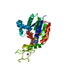
| ||||||||
|---|---|---|---|---|---|---|---|---|---|
| 1 |
| ||||||||
| Unit cell |
|
-Components
-Protein , 1 types, 1 molecules A
| #1: Protein | Mass: 25599.391 Da / Num. of mol.: 1 Source method: isolated from a genetically manipulated source Source: (gene. exp.) Homo sapiens (human) / Gene: AK3, AK3L1, AK6, AKL3L / Production host:  Escherichia coli (E. coli) Escherichia coli (E. coli)References: UniProt: Q9UIJ7, nucleoside-triphosphate-adenylate kinase |
|---|
-Non-polymers , 5 types, 313 molecules 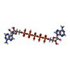


| #2: Chemical | ChemComp-AP5 /  Mass: 916.367 Da / Num. of mol.: 1 / Source method: obtained synthetically / Formula: C20H29N10O22P5 / Feature type: SUBJECT OF INVESTIGATION Mass: 916.367 Da / Num. of mol.: 1 / Source method: obtained synthetically / Formula: C20H29N10O22P5 / Feature type: SUBJECT OF INVESTIGATION | ||||||
|---|---|---|---|---|---|---|---|
| #3: Chemical | Chloride Mass: 35.453 Da / Num. of mol.: 2 / Source method: obtained synthetically / Formula: Cl Mass: 35.453 Da / Num. of mol.: 2 / Source method: obtained synthetically / Formula: Cl#4: Chemical | ChemComp-NA / |  Mass: 22.990 Da / Num. of mol.: 1 / Source method: obtained synthetically / Formula: Na Mass: 22.990 Da / Num. of mol.: 1 / Source method: obtained synthetically / Formula: Na#5: Chemical | ChemComp-MG / |  Mass: 24.305 Da / Num. of mol.: 1 / Source method: obtained synthetically / Formula: Mg Mass: 24.305 Da / Num. of mol.: 1 / Source method: obtained synthetically / Formula: Mg#6: Water | ChemComp-HOH / | WaterMass: 18.015 Da / Num. of mol.: 308 / Source method: isolated from a natural source / Formula: H2O |
-Details
| Has ligand of interest | Y |
|---|
-Experimental details
-Experiment
| Experiment | Method: X-RAY DIFFRACTION / Number of used crystals: 1 |
|---|
- Sample preparation
| Crystal | Density Matthews: 2.49 Å3/Da / Density % sol: 50.66 % |
|---|---|
| Crystal grow | Temperature: 291 K / Method: vapor diffusion, hanging drop / pH: 8 Details: Purified hAK3 was dialyzed against 30 mM MOPS buffer with 50 mM NaCl pH 7.0 and concentrated to 15-20 mg per ml. The resevoir contained 0.2 M CaCl2, 0.1 M Tris pH 8.0 and 20% PEG 6000. Drop ...Details: Purified hAK3 was dialyzed against 30 mM MOPS buffer with 50 mM NaCl pH 7.0 and concentrated to 15-20 mg per ml. The resevoir contained 0.2 M CaCl2, 0.1 M Tris pH 8.0 and 20% PEG 6000. Drop size 1 plus 1 microliter. Cryo protection 30% PEG 6000 |
-Data collection
| Diffraction | Mean temperature: 100 K / Serial crystal experiment: N |
|---|---|
| Diffraction source | Source: SYNCHROTRON / Site: ESRF  / Beamline: ID23-2 / Wavelength: 0.873 Å / Beamline: ID23-2 / Wavelength: 0.873 Å |
| Detector | Type: DECTRIS PILATUS 6M-F / Detector: PIXEL / Date: Oct 24, 2018 |
| Radiation | Protocol: SINGLE WAVELENGTH / Monochromatic (M) / Laue (L): M / Scattering type: x-ray |
| Radiation wavelength | Wavelength: 0.873 Å / Relative weight: 1 |
| Reflection | Resolution: 1.48→42.72 Å / Num. obs: 39892 / % possible obs: 95.1 % / Redundancy: 6 % / CC1/2: 0.999 / Rmerge(I) obs: 0.05 / Rpim(I) all: 0.033 / Net I/σ(I): 14.9 |
| Reflection shell | Resolution: 1.48→1.53 Å / Redundancy: 4.3 % / Rmerge(I) obs: 0.852 / Mean I/σ(I) obs: 1.3 / Num. unique obs: 3174 / CC1/2: 0.521 / Rpim(I) all: 0.707 / % possible all: 76.1 |
- Processing
| Software |
| ||||||||||||||||||||||||||||||||||||||||||||||||||||||||||||||||||||||||||||||||||||||||||
|---|---|---|---|---|---|---|---|---|---|---|---|---|---|---|---|---|---|---|---|---|---|---|---|---|---|---|---|---|---|---|---|---|---|---|---|---|---|---|---|---|---|---|---|---|---|---|---|---|---|---|---|---|---|---|---|---|---|---|---|---|---|---|---|---|---|---|---|---|---|---|---|---|---|---|---|---|---|---|---|---|---|---|---|---|---|---|---|---|---|---|---|
| Refinement | Method to determine structure: MOLECULAR REPLACEMENT Starting model: 1zd8 Resolution: 1.48→40.624 Å / SU ML: 0.15 / Cross valid method: THROUGHOUT / σ(F): 1.38 / Phase error: 19.29 / Stereochemistry target values: ML
| ||||||||||||||||||||||||||||||||||||||||||||||||||||||||||||||||||||||||||||||||||||||||||
| Solvent computation | Shrinkage radii: 0.9 Å / VDW probe radii: 1.11 Å / Solvent model: FLAT BULK SOLVENT MODEL | ||||||||||||||||||||||||||||||||||||||||||||||||||||||||||||||||||||||||||||||||||||||||||
| Displacement parameters | Biso max: 106.64 Å2 / Biso mean: 30.4604 Å2 / Biso min: 15.27 Å2 | ||||||||||||||||||||||||||||||||||||||||||||||||||||||||||||||||||||||||||||||||||||||||||
| Refinement step | Cycle: final / Resolution: 1.48→40.624 Å
| ||||||||||||||||||||||||||||||||||||||||||||||||||||||||||||||||||||||||||||||||||||||||||
| LS refinement shell | Refine-ID: X-RAY DIFFRACTION / Rfactor Rfree error: 0
|
