 Movie
Movie Controller

[English] 日本語
 Yorodumi
- PDB-6zj5: Structure of the catalytic domain of human endo-alpha-mannosidase... -
+ Open data

- Basic information
| Entry | Database: PDB / ID: 6zj5 | |||||||||||||||
|---|---|---|---|---|---|---|---|---|---|---|---|---|---|---|---|---|
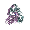
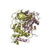
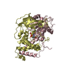
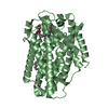
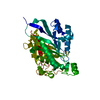
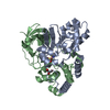
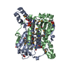
| Title | Structure of the catalytic domain of human endo-alpha-mannosidase MANEA in complex with GlcDMJ and hexatungstotellurate(VI) TEW | |||||||||||||||
 Components Components | Glycoprotein endo-alpha-1,2-mannosidase | |||||||||||||||
 Keywords Keywords | HYDROLASE / Golgi / mannosidase / retaining | |||||||||||||||
| Function / homology |  Function and homology informationglycoprotein endo-alpha-1,2-mannosidase / glycoprotein endo-alpha-1,2-mannosidase activity / N-glycan trimming and elongation in the cis-Golgi / alpha-mannosidase activity / Golgi membrane / Golgi apparatus Function and homology informationglycoprotein endo-alpha-1,2-mannosidase / glycoprotein endo-alpha-1,2-mannosidase activity / N-glycan trimming and elongation in the cis-Golgi / alpha-mannosidase activity / Golgi membrane / Golgi apparatusSimilarity search - Function | |||||||||||||||
| Biological species |  Homo sapiens (human) Homo sapiens (human) | |||||||||||||||
| Method | X-RAY DIFFRACTION / SYNCHROTRON / MOLECULAR REPLACEMENT / Resolution: 2.269 Å | |||||||||||||||
 Authors Authors | Sobala, L.F. / Fernandes, P.Z. / Hakki, Z. / Thompson, A.J. / Howe, J.D. / Hill, M. / Zitzmann, N. / Davies, S. / Stamataki, Z. / Butters, T.D. ...Sobala, L.F. / Fernandes, P.Z. / Hakki, Z. / Thompson, A.J. / Howe, J.D. / Hill, M. / Zitzmann, N. / Davies, S. / Stamataki, Z. / Butters, T.D. / Alonzi, D.S. / Williams, S.J. / Davies, G.J. | |||||||||||||||
| Funding support |  United Kingdom, United Kingdom,  Australia, 4items Australia, 4items
| |||||||||||||||
 Citation Citation | Journal: Proc.Natl.Acad.Sci.USA / Year: 2020 Title: Structure of human endo-alpha-1,2-mannosidase (MANEA), an antiviral host-glycosylation target. Authors: Sobala, L.F. / Fernandes, P.Z. / Hakki, Z. / Thompson, A.J. / Howe, J.D. / Hill, M. / Zitzmann, N. / Davies, S. / Stamataki, Z. / Butters, T.D. / Alonzi, D.S. / Williams, S.J. / Davies, G.J. | |||||||||||||||
| History |
|
- Structure visualization
| Structure viewer | Molecule: MolmilJmol/JSmol |
|---|
- Downloads & links
-Download
| PDBx/mmCIF format | 6zj5.cif.gz | 101.2 KB | Display | PDBx/mmCIF format |
|---|---|---|---|---|
| PDB format | pdb6zj5.ent.gz | Display | PDB format | |
| PDBx/mmJSON format | 6zj5.json.gz | Tree view | PDBx/mmJSON format | |
| Others |  Other downloads Other downloads |
-Validation report
| Arichive directory | https://data.pdbj.org/pub/pdb/validation_reports/zj/6zj5ftp://data.pdbj.org/pub/pdb/validation_reports/zj/6zj5 | HTTPS FTP |
|---|
-Related structure data
| Related structure data |  6zdcC  6zdfC  6zdkC  6zdlC  6zfaSC  6zfnC  6zfqC  6zj1C 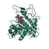 6zj6C S: Starting model for refinement C: citing same article ( |
|---|---|
| Similar structure data | |
| Experimental dataset #1 | Data reference: 10.5281/zenodo.4288341 / Data set type: diffraction image data |


-Links
 PDBj
PDBj

- Assembly
| Deposited unit | 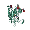
| ||||||||
|---|---|---|---|---|---|---|---|---|---|
| 1 |
| ||||||||
| Unit cell |
| ||||||||
| Components on special symmetry positions |
|
-Components
-Protein / Sugars , 2 types, 2 molecules AAA
| #1: Protein | / hEndo / Mandaselin Mass: 44783.094 Da / Num. of mol.: 1 Source method: isolated from a genetically manipulated source Source: (gene. exp.) Homo sapiens (human) / Gene: MANEA / Plasmid: pCold-I / Production host:  Escherichia coli BL21(DE3) (bacteria) Escherichia coli BL21(DE3) (bacteria)References: UniProt: Q5SRI9, glycoprotein endo-alpha-1,2-mannosidase |
|---|---|
| #6: Sugar | ChemComp-GLC / Glucose Type: D-saccharide, alpha linking / Mass: 180.156 Da / Num. of mol.: 1 Type: D-saccharide, alpha linking / Mass: 180.156 Da / Num. of mol.: 1Source method: isolated from a genetically manipulated source Formula: C6H12O6 / Feature type: SUBJECT OF INVESTIGATION |
-Non-polymers , 5 types, 102 molecules 


| #2: Chemical | HEPES Mass: 238.305 Da / Num. of mol.: 3 / Source method: obtained synthetically / Formula: C8H18N2O4S / Comment: pH buffer*YM Mass: 238.305 Da / Num. of mol.: 3 / Source method: obtained synthetically / Formula: C8H18N2O4S / Comment: pH buffer*YM#3: Chemical | ChemComp-MG / |  Mass: 24.305 Da / Num. of mol.: 1 / Source method: obtained synthetically / Formula: Mg Mass: 24.305 Da / Num. of mol.: 1 / Source method: obtained synthetically / Formula: Mg#4: Chemical |  Mass: 1614.626 Da / Num. of mol.: 3 / Source method: obtained synthetically / Formula: O24TeW6 Mass: 1614.626 Da / Num. of mol.: 3 / Source method: obtained synthetically / Formula: O24TeW6#5: Chemical | ChemComp-DMJ / |  Mass: 163.172 Da / Num. of mol.: 1 / Source method: obtained synthetically / Formula: C6H13NO4 / Feature type: SUBJECT OF INVESTIGATION Mass: 163.172 Da / Num. of mol.: 1 / Source method: obtained synthetically / Formula: C6H13NO4 / Feature type: SUBJECT OF INVESTIGATION#7: Water | ChemComp-HOH / | WaterMass: 18.015 Da / Num. of mol.: 94 / Source method: isolated from a natural source / Formula: H2O |
|---|
-Details
| Has ligand of interest | Y |
|---|
-Experimental details
-Experiment
| Experiment | Method: X-RAY DIFFRACTION / Number of used crystals: 1 |
|---|
- Sample preparation
| Crystal | Density Matthews: 2.57 Å3/Da / Density % sol: 52.2 % / Description: hexagonal, flat |
|---|---|
| Crystal grow | Temperature: 292 K / Method: vapor diffusion, sitting drop Details: 100 mM HEPES pH 7.5 - 8.1, 200 mM MgCl2, 25-27.5% v/v PEG 400, 1 mM TEW Protein in 25 mM HEPES pH 7.0, 200 mM NaCl buffer at 10 mg/ml. PH range: 7.5-8.1 |
-Data collection
| Diffraction | Mean temperature: 100 K / Serial crystal experiment: N |
|---|---|
| Diffraction source | Source: SYNCHROTRON / Site: Diamond / Beamline: I04 / Wavelength: 0.97949 Å |
| Detector | Type: DECTRIS EIGER2 XE 16M / Detector: PIXEL / Date: Mar 7, 2019 |
| Radiation | Protocol: SINGLE WAVELENGTH / Monochromatic (M) / Laue (L): M / Scattering type: x-ray |
| Radiation wavelength | Wavelength: 0.97949 Å / Relative weight: 1 |
| Reflection | Resolution: 2.269→111.327 Å / Num. obs: 11089 / % possible obs: 51.9 % / Redundancy: 8 % / CC1/2: 0.989 / Rmerge(I) obs: 0.276 / Rpim(I) all: 0.103 / Rrim(I) all: 0.295 / Net I/σ(I): 5.3 |
| Reflection shell | Resolution: 2.269→2.536 Å / Redundancy: 8.6 % / Rmerge(I) obs: 1.52 / Num. unique obs: 925 / CC1/2: 0.569 / Rpim(I) all: 0.547 / Rrim(I) all: 1.617 / % possible all: 15.4 |
- Processing
| Software |
| |||||||||||||||||||||||||||||||||||||||||||||||||||||||||||||||||||||||||||||||||||||||||||||||||||||||||||||||||||||||||||||||||||||||||||||||||||||||||||||||||||||||||||||||||||||||||||||||||||||||||||||||||||||||||||||||||||||||
|---|---|---|---|---|---|---|---|---|---|---|---|---|---|---|---|---|---|---|---|---|---|---|---|---|---|---|---|---|---|---|---|---|---|---|---|---|---|---|---|---|---|---|---|---|---|---|---|---|---|---|---|---|---|---|---|---|---|---|---|---|---|---|---|---|---|---|---|---|---|---|---|---|---|---|---|---|---|---|---|---|---|---|---|---|---|---|---|---|---|---|---|---|---|---|---|---|---|---|---|---|---|---|---|---|---|---|---|---|---|---|---|---|---|---|---|---|---|---|---|---|---|---|---|---|---|---|---|---|---|---|---|---|---|---|---|---|---|---|---|---|---|---|---|---|---|---|---|---|---|---|---|---|---|---|---|---|---|---|---|---|---|---|---|---|---|---|---|---|---|---|---|---|---|---|---|---|---|---|---|---|---|---|---|---|---|---|---|---|---|---|---|---|---|---|---|---|---|---|---|---|---|---|---|---|---|---|---|---|---|---|---|---|---|---|---|---|---|---|---|---|---|---|---|---|---|---|---|---|---|---|---|---|
| Refinement | Method to determine structure: MOLECULAR REPLACEMENT Starting model: 6ZFA Resolution: 2.269→111.327 Å / Cor.coef. Fo:Fc: 0.946 / Cor.coef. Fo:Fc free: 0.895 / WRfactor Rfree: 0.241 / WRfactor Rwork: 0.174 / SU B: 10.303 / SU ML: 0.246 / Average fsc free: 0.8681 / Average fsc work: 0.8925 / Cross valid method: FREE R-VALUE / ESU R Free: 0.408 Details: Hydrogens have been added in their riding positions
| |||||||||||||||||||||||||||||||||||||||||||||||||||||||||||||||||||||||||||||||||||||||||||||||||||||||||||||||||||||||||||||||||||||||||||||||||||||||||||||||||||||||||||||||||||||||||||||||||||||||||||||||||||||||||||||||||||||||
| Solvent computation | Ion probe radii: 0.8 Å / Shrinkage radii: 0.8 Å / VDW probe radii: 1.2 Å / Solvent model: MASK BULK SOLVENT | |||||||||||||||||||||||||||||||||||||||||||||||||||||||||||||||||||||||||||||||||||||||||||||||||||||||||||||||||||||||||||||||||||||||||||||||||||||||||||||||||||||||||||||||||||||||||||||||||||||||||||||||||||||||||||||||||||||||
| Displacement parameters | Biso mean: 29.85 Å2
| |||||||||||||||||||||||||||||||||||||||||||||||||||||||||||||||||||||||||||||||||||||||||||||||||||||||||||||||||||||||||||||||||||||||||||||||||||||||||||||||||||||||||||||||||||||||||||||||||||||||||||||||||||||||||||||||||||||||
| Refinement step | Cycle: LAST / Resolution: 2.269→111.327 Å
| |||||||||||||||||||||||||||||||||||||||||||||||||||||||||||||||||||||||||||||||||||||||||||||||||||||||||||||||||||||||||||||||||||||||||||||||||||||||||||||||||||||||||||||||||||||||||||||||||||||||||||||||||||||||||||||||||||||||
| Refine LS restraints |
| |||||||||||||||||||||||||||||||||||||||||||||||||||||||||||||||||||||||||||||||||||||||||||||||||||||||||||||||||||||||||||||||||||||||||||||||||||||||||||||||||||||||||||||||||||||||||||||||||||||||||||||||||||||||||||||||||||||||
| LS refinement shell | Refine-ID: X-RAY DIFFRACTION / Total num. of bins used: 20
|
