 Movie
Movie Controller


+ Open data

- Basic information
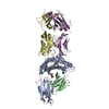
| Entry | Database: PDB / ID: 6z8h | ||||||
|---|---|---|---|---|---|---|---|
| Title | Crystal structure of Variant Surface Glycoprotein VSG13 | ||||||
 Components Components | Variant surface glycoprotein MITat 1.13 | ||||||
 Keywords Keywords |  MEMBRANE PROTEIN / Variant Surface Glycoprotein / Immune Recognition / Immune Evasion / Trypanosomiasis MEMBRANE PROTEIN / Variant Surface Glycoprotein / Immune Recognition / Immune Evasion / Trypanosomiasis | ||||||
| Function / homology | Trypanosome variant surface glycoprotein, C-terminal / Trypanosome variant surface glycoprotein C-terminal domain / Trypanosome variant surface glycoprotein, A-type, N-terminal domain / Trypanosome variant surface glycoprotein (A-type) / evasion of host immune response / plasma membrane / Variant surface glycoprotein MITat 1.13 Function and homology information Function and homology information | ||||||
| Biological species |  Trypanosoma brucei (eukaryote) Trypanosoma brucei (eukaryote) | ||||||
| Method | X-RAY DIFFRACTION / SYNCHROTRON / MOLECULAR REPLACEMENT / Resolution: 1.38 Å | ||||||
 Authors Authors | Stebbins, C.E. / Hempelmann, A. / Van Straaten, M. / Zeelen, J. | ||||||
 Citation Citation | Journal: Nat Microbiol / Year: 2021 Title: Structure of trypanosome coat protein VSGsur and function in suramin resistance. Authors: Zeelen, J. / van Straaten, M. / Verdi, J. / Hempelmann, A. / Hashemi, H. / Perez, K. / Jeffrey, P.D. / Halg, S. / Wiedemar, N. / Maser, P. / Papavasiliou, F.N. / Stebbins, C.E. | ||||||
| History |
|
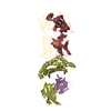
- Structure visualization
| Structure viewer | Molecule: MolmilJmol/JSmol |
|---|
- Downloads & links
-Download
| PDBx/mmCIF format | 6z8h.cif.gz | 441.9 KB | Display | PDBx/mmCIF format |
|---|---|---|---|---|
| PDB format | pdb6z8h.ent.gz | 335.1 KB | Display | PDB format |
| PDBx/mmJSON format | 6z8h.json.gz | Tree view | PDBx/mmJSON format | |
| Others |  Other downloads Other downloads |
-Validation report
| Arichive directory | https://data.pdbj.org/pub/pdb/validation_reports/z8/6z8hftp://data.pdbj.org/pub/pdb/validation_reports/z8/6z8h | HTTPS FTP |
|---|
-Related structure data
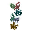
| Related structure data |  6z79C 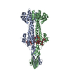 6z7bC  6z7cC  6z7dC  6z7eC 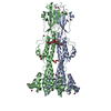 6z8gSC S: Starting model for refinement C: citing same article ( |
|---|---|
| Similar structure data |


-Links
 PDBj
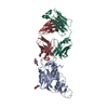
PDBj- Assembly
| Deposited unit | 
| ||||||||||||
|---|---|---|---|---|---|---|---|---|---|---|---|---|---|
| 1 |
| ||||||||||||
| Unit cell |
|
-Components
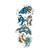
| #1: Protein | Mass: 53771.410 Da / Num. of mol.: 2 / Source method: isolated from a natural source / Details: Glycosylated at N260 / Source: (natural) Trypanosoma brucei (eukaryote) / References: UniProt: Q58NS4#2: Polysaccharide | / Mass: 342.297 Da / Num. of mol.: 2Source method: isolated from a genetically manipulated source #3: Sugar | ChemComp-NAG / N-Acetylglucosamine  Type: D-saccharide, beta linking / Mass: 221.208 Da / Num. of mol.: 4 Type: D-saccharide, beta linking / Mass: 221.208 Da / Num. of mol.: 4Source method: isolated from a genetically manipulated source Formula: C8H15NO6 #4: Chemical | ChemComp-SO4 / | Sulfate  Mass: 96.063 Da / Num. of mol.: 1 Mass: 96.063 Da / Num. of mol.: 1Source method: isolated from a genetically manipulated source Formula: SO4 #5: Water | ChemComp-HOH / | Water Mass: 18.015 Da / Num. of mol.: 421 / Source method: isolated from a natural source / Formula: H2O Mass: 18.015 Da / Num. of mol.: 421 / Source method: isolated from a natural source / Formula: H2OHas ligand of interest | N | |
|---|
-Experimental details
-Experiment
| Experiment | Method: X-RAY DIFFRACTION / Number of used crystals: 1 |
|---|
- Sample preparation
| Crystal grow | Temperature: 296 K / Method: vapor diffusion, hanging drop / pH: 8.5 / Details: 1.8-2.0 M (NH4)2SO4, 100 mM Tris.Cl pH 8.5 |
|---|
-Data collection
| Diffraction | Mean temperature: 100 K / Serial crystal experiment: N |
|---|---|
| Diffraction source | Source: SYNCHROTRON / Site: ESRF  / Beamline: ID29 / Wavelength: 1 Å / Beamline: ID29 / Wavelength: 1 Å |
| Detector | Type: DECTRIS PILATUS 6M-F / Detector: PIXEL / Date: Apr 12, 2018 |
| Radiation | Protocol: SINGLE WAVELENGTH / Monochromatic (M) / Laue (L): M / Scattering type: x-ray |
| Radiation wavelength | Wavelength: 1 Å / Relative weight: 1 |
| Reflection | Resolution: 1.378→48.14 Å / Num. obs: 155980 / % possible obs: 97.09 % / Redundancy: 3 % / Biso Wilson estimate: 18.5 Å2 / CC1/2: 0.995 / CC star: 0.999 / Rpim(I) all: 0.03368 / Net I/σ(I): 8.07 |
| Reflection shell | Resolution: 1.378→1.428 Å / Num. unique obs: 15141 / CC1/2: 0.84 / % possible all: 94.49 |
- Processing
| Software |
| |||||||||||||||||||||||||||||||||||||||||||||||||||||||||||||||||||||||||||||||||||||||||||
|---|---|---|---|---|---|---|---|---|---|---|---|---|---|---|---|---|---|---|---|---|---|---|---|---|---|---|---|---|---|---|---|---|---|---|---|---|---|---|---|---|---|---|---|---|---|---|---|---|---|---|---|---|---|---|---|---|---|---|---|---|---|---|---|---|---|---|---|---|---|---|---|---|---|---|---|---|---|---|---|---|---|---|---|---|---|---|---|---|---|---|---|---|
| Refinement | Method to determine structure: MOLECULAR REPLACEMENT Starting model: 6Z8G Resolution: 1.38→48.14 Å / SU ML: 0.1897 / Cross valid method: THROUGHOUT / σ(F): 1.34 / Phase error: 32.7523 Stereochemistry target values: GeoStd + Monomer Library + CDL v1.2
| |||||||||||||||||||||||||||||||||||||||||||||||||||||||||||||||||||||||||||||||||||||||||||
| Solvent computation | Shrinkage radii: 0.9 Å / VDW probe radii: 1.11 Å / Solvent model: FLAT BULK SOLVENT MODEL | |||||||||||||||||||||||||||||||||||||||||||||||||||||||||||||||||||||||||||||||||||||||||||
| Displacement parameters | Biso mean: 31.18 Å2 | |||||||||||||||||||||||||||||||||||||||||||||||||||||||||||||||||||||||||||||||||||||||||||
| Refinement step | Cycle: LAST / Resolution: 1.38→48.14 Å
| |||||||||||||||||||||||||||||||||||||||||||||||||||||||||||||||||||||||||||||||||||||||||||
| Refine LS restraints |
| |||||||||||||||||||||||||||||||||||||||||||||||||||||||||||||||||||||||||||||||||||||||||||
| LS refinement shell |
|
