 Movie
Movie Controller

[English] 日本語
 Yorodumi
- PDB-6z63: FtsE structure from Streptococus pneumoniae in complex with ADP a... -
+ Open data

- Basic information
| Entry | Database: PDB / ID: 6z63 | ||||||
|---|---|---|---|---|---|---|---|
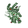
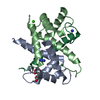
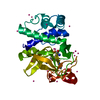
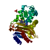
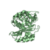
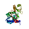
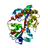
| Title | FtsE structure from Streptococus pneumoniae in complex with ADP at 1.57 A resolution (spacegroup P 21) | ||||||
 Components Components | Cell division ATP-binding protein FtsE | ||||||
 Keywords Keywords |  CELL CYCLE / Cell division / divisome / FtsEX / ATP-binding protein CELL CYCLE / Cell division / divisome / FtsEX / ATP-binding protein | ||||||
| Function / homology |  Function and homology information Function and homology informationtransmembrane transporter activity / transmembrane transport / cell cycle / cell division / ATP hydrolysis activity / ATP binding / plasma membraneSimilarity search - Function | ||||||
| Biological species |   Streptococcus pneumoniae (bacteria) Streptococcus pneumoniae (bacteria) | ||||||
| Method | X-RAY DIFFRACTION / SYNCHROTRON / MOLECULAR REPLACEMENT / Resolution: 1.57 Å | ||||||
 Authors Authors | Alcorlo, M. / Straume, D. / Havarstein, L.S. / Hermoso, J.A. | ||||||
| Funding support |  Spain, 1items Spain, 1items
| ||||||
 Citation Citation | Journal: Mbio / Year: 2020 Title: Structural Characterization of the Essential Cell Division Protein FtsE and Its Interaction with FtsX in Streptococcus pneumoniae. Authors: Alcorlo, M. / Straume, D. / Lutkenhaus, J. / Havarstein, L.S. / Hermoso, J.A. | ||||||
| History |
|
- Structure visualization
| Structure viewer | Molecule: MolmilJmol/JSmol |
|---|
- Downloads & links
-Download
| PDBx/mmCIF format | 6z63.cif.gz | 167.1 KB | Display | PDBx/mmCIF format |
|---|---|---|---|---|
| PDB format | pdb6z63.ent.gz | 131.1 KB | Display | PDB format |
| PDBx/mmJSON format | 6z63.json.gz | Tree view | PDBx/mmJSON format | |
| Others |  Other downloads Other downloads |
-Validation report
| Arichive directory | https://data.pdbj.org/pub/pdb/validation_reports/z6/6z63ftp://data.pdbj.org/pub/pdb/validation_reports/z6/6z63 | HTTPS FTP |
|---|
-Related structure data
| Related structure data | 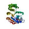 6z4wC  6z67C  2oukS S: Starting model for refinement C: citing same article ( |
|---|---|
| Similar structure data |


-Links
 PDBj
PDBj

- Assembly
| Deposited unit | 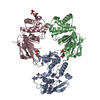
| ||||||||
|---|---|---|---|---|---|---|---|---|---|
| 1 | 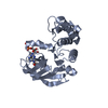
| ||||||||
| 2 | 
| ||||||||
| 3 | 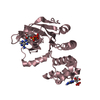
| ||||||||
| Unit cell |
|
-Components
| #1: Protein | Mass: 25760.676 Da / Num. of mol.: 3 Source method: isolated from a genetically manipulated source Source: (gene. exp.) Streptococcus pneumoniae (bacteria)Gene: ftsE_1, ftsE, ftsE_2, AZJ28_05890, AZJ96_05755, AZK21_09500, AZK39_08795, CWI64_10710, ERS003549_00229, ERS019159_00859, ERS019166_00608, ERS019258_00545, ERS019260_01795, ERS019499_01745, ...Gene: ftsE_1, ftsE, ftsE_2, AZJ28_05890, AZJ96_05755, AZK21_09500, AZK39_08795, CWI64_10710, ERS003549_00229, ERS019159_00859, ERS019166_00608, ERS019258_00545, ERS019260_01795, ERS019499_01745, ERS020087_02007, ERS020408_00564, ERS020474_01891, ERS020873_00605, ERS021072_00923, ERS021218_01619, ERS021243_01290, ERS043879_00383, ERS050419_00449, ERS232443_01143, ERS232484_01031, ERS368006_01003, ERS409062_02036, ERS409277_00646, NCTC12140_01748, SAMEA104035134_02097, SAMEA104035170_01887, SAMEA104154666_00013, SAMEA2052026_00651, SAMEA2203388_01270, SAMEA2203858_01390, SAMEA2335963_01162, SAMEA2335976_01110, SAMEA2341322_01915, SAMEA2521606_00082, SAMEA2521861_00196, SAMEA2696310_01884, SAMEA2696394_01822, SAMEA2696492_00847, SAMEA2696596_00644, SAMEA2796717_01437, SAMEA2796719_00990, SAMEA3171064_02197, SAMEA3172940_00439, SAMEA3173021_00704, SAMEA3207192_00670, SAMEA3207204_00699, SAMEA3232645_02031, SAMEA3309623_00023, SAMEA3353431_01338, SAMEA3353605_01219, SAMEA3353631_01406, SAMEA3354309_00922, SAMEA3381574_01396, SAMEA3389847_01128, SAMEA3390019_01763, SAMEA3506052_00474, SAMEA3714202_02061, SAMEA3714261_01293, SAMEA3714340_00855, SAMEA4038883_00064, SAMEA4388199_00912, SpnNT_00739 Production host: Escherichia coli (E. coli) / References: UniProt: A0A064BZ20, UniProt: Q8DQH4*PLUS#2: Chemical | ChemComp-ADP / Adenosine diphosphate  Mass: 427.201 Da / Num. of mol.: 5 / Source method: obtained synthetically / Formula: C10H15N5O10P2 / Feature type: SUBJECT OF INVESTIGATION / Comment: ADP, energy-carrying molecule*YM Mass: 427.201 Da / Num. of mol.: 5 / Source method: obtained synthetically / Formula: C10H15N5O10P2 / Feature type: SUBJECT OF INVESTIGATION / Comment: ADP, energy-carrying molecule*YM#3: Water | ChemComp-HOH / | Water Mass: 18.015 Da / Num. of mol.: 645 / Source method: isolated from a natural source / Formula: H2O Mass: 18.015 Da / Num. of mol.: 645 / Source method: isolated from a natural source / Formula: H2OHas ligand of interest | Y | |
|---|
-Experimental details
-Experiment
| Experiment | Method: X-RAY DIFFRACTION / Number of used crystals: 1 |
|---|
- Sample preparation
| Crystal | Density Matthews: 2.06 Å3/Da / Density % sol: 40.4 % |
|---|---|
| Crystal grow | Temperature: 291 K / Method: vapor diffusion, sitting drop / Details: 0.15 M NaF and 16% (w/v) PEG3350 |
-Data collection
| Diffraction | Mean temperature: 100 K / Serial crystal experiment: N |
|---|---|
| Diffraction source | Source: SYNCHROTRON / Site: ALBA / Beamline: XALOC / Wavelength: 0.96862 Å |
| Detector | Type: DECTRIS PILATUS3 S 6M / Detector: PIXEL / Date: Apr 17, 2017 |
| Radiation | Protocol: SINGLE WAVELENGTH / Monochromatic (M) / Laue (L): M / Scattering type: x-ray |
| Radiation wavelength | Wavelength: 0.96862 Å / Relative weight: 1 |
| Reflection | Resolution: 1.57→47.97 Å / Num. obs: 86937 / % possible obs: 99.5 % / Redundancy: 5 % / CC1/2: 0.99 / Rmerge(I) obs: 0.081 / Net I/σ(I): 11.7 |
| Reflection shell | Resolution: 1.57→1.6 Å / Rmerge(I) obs: 0.817 / Num. unique obs: 4252 / CC1/2: 0.56 |
- Processing
| Software |
| ||||||||||||||||||||||||||||||||||||||||||||||||||||||||||||||||||||||||||||||||||||||||||||||||||||||||||||||||||||||||||||||||||||||||||||||||||||||||||||||||||||||||||||||||||||||||||
|---|---|---|---|---|---|---|---|---|---|---|---|---|---|---|---|---|---|---|---|---|---|---|---|---|---|---|---|---|---|---|---|---|---|---|---|---|---|---|---|---|---|---|---|---|---|---|---|---|---|---|---|---|---|---|---|---|---|---|---|---|---|---|---|---|---|---|---|---|---|---|---|---|---|---|---|---|---|---|---|---|---|---|---|---|---|---|---|---|---|---|---|---|---|---|---|---|---|---|---|---|---|---|---|---|---|---|---|---|---|---|---|---|---|---|---|---|---|---|---|---|---|---|---|---|---|---|---|---|---|---|---|---|---|---|---|---|---|---|---|---|---|---|---|---|---|---|---|---|---|---|---|---|---|---|---|---|---|---|---|---|---|---|---|---|---|---|---|---|---|---|---|---|---|---|---|---|---|---|---|---|---|---|---|---|---|---|---|
| Refinement | Method to determine structure: MOLECULAR REPLACEMENT Starting model: 2OUK Resolution: 1.57→47.967 Å / SU ML: 0.19 / Cross valid method: THROUGHOUT / σ(F): 1.35 / Phase error: 22.33 / Stereochemistry target values: ML
| ||||||||||||||||||||||||||||||||||||||||||||||||||||||||||||||||||||||||||||||||||||||||||||||||||||||||||||||||||||||||||||||||||||||||||||||||||||||||||||||||||||||||||||||||||||||||||
| Solvent computation | Shrinkage radii: 0.9 Å / VDW probe radii: 1.11 Å / Solvent model: FLAT BULK SOLVENT MODEL | ||||||||||||||||||||||||||||||||||||||||||||||||||||||||||||||||||||||||||||||||||||||||||||||||||||||||||||||||||||||||||||||||||||||||||||||||||||||||||||||||||||||||||||||||||||||||||
| Displacement parameters | Biso max: 108.27 Å2 / Biso mean: 22.972 Å2 / Biso min: 3.1 Å2 | ||||||||||||||||||||||||||||||||||||||||||||||||||||||||||||||||||||||||||||||||||||||||||||||||||||||||||||||||||||||||||||||||||||||||||||||||||||||||||||||||||||||||||||||||||||||||||
| Refinement step | Cycle: final / Resolution: 1.57→47.967 Å
| ||||||||||||||||||||||||||||||||||||||||||||||||||||||||||||||||||||||||||||||||||||||||||||||||||||||||||||||||||||||||||||||||||||||||||||||||||||||||||||||||||||||||||||||||||||||||||
| LS refinement shell | Refine-ID: X-RAY DIFFRACTION / Rfactor Rfree error: 0
|
