 Movie
Movie Controller

[English] 日本語
 Yorodumi
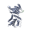
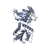
- PDB-6z3j: Repulsive Guidance Molecule B (RGMB) in complex with Growth Diffe... -
+ Open data

- Basic information
| Entry | Database: PDB / ID: 6z3j | ||||||||||||||||||||||||
|---|---|---|---|---|---|---|---|---|---|---|---|---|---|---|---|---|---|---|---|---|---|---|---|---|---|
| Title | Repulsive Guidance Molecule B (RGMB) in complex with Growth Differentiation Factor 5 (GDF5) (crystal form 1) | ||||||||||||||||||||||||
 Components Components |
| ||||||||||||||||||||||||
 Keywords Keywords |  SIGNALING PROTEIN / Repulsive Guidance Molecule / RGM / Bone Morphogenetic Protein / BMP / Growth Differentiation Factor 5 / GDF5 / Neogenin / axon guidance / TGFbeta signalling / brain development / iron metabolism. SIGNALING PROTEIN / Repulsive Guidance Molecule / RGM / Bone Morphogenetic Protein / BMP / Growth Differentiation Factor 5 / GDF5 / Neogenin / axon guidance / TGFbeta signalling / brain development / iron metabolism. | ||||||||||||||||||||||||
| Function / homology |  Function and homology information Function and homology informationossification involved in bone remodeling / forelimb morphogenesis / BMP binding / chondroblast differentiation / hindlimb morphogenesis / negative regulation of mesenchymal cell apoptotic process / Netrin-1 signaling / positive regulation of chondrocyte differentiation / mesenchymal cell apoptotic process / positive regulation of BMP signaling pathway ...ossification involved in bone remodeling / forelimb morphogenesis / BMP binding / chondroblast differentiation / hindlimb morphogenesis / negative regulation of mesenchymal cell apoptotic process / Netrin-1 signaling / positive regulation of chondrocyte differentiation / mesenchymal cell apoptotic process / positive regulation of BMP signaling pathway / negative regulation of chondrocyte differentiation / embryonic limb morphogenesis / Molecules associated with elastic fibres / positive regulation of SMAD protein signal transduction / endoplasmic reticulum-Golgi intermediate compartment / regulation of multicellular organism growth / chondrocyte differentiation / BMP signaling pathway / coreceptor activity / side of membrane / response to mechanical stimulus / positive regulation of neuron differentiation / transforming growth factor beta receptor signaling pathway / cytokine activity / growth factor activity / negative regulation of epithelial cell proliferation / cell-cell signaling / negative regulation of neuron apoptotic process / cell adhesion / membrane raft / positive regulation of DNA-templated transcription / signal transduction / extracellular space / extracellular region / identical protein binding / plasma membraneSimilarity search - Function | ||||||||||||||||||||||||
| Biological species |  Homo sapiens (human) Homo sapiens (human) | ||||||||||||||||||||||||
| Method | X-RAY DIFFRACTION / SYNCHROTRON / MOLECULAR REPLACEMENT / Resolution: 1.65 Å | ||||||||||||||||||||||||
 Authors Authors | Malinauskas, T. / Peer, T.V. / Bishop, B. / Muller, T.D. / Siebold, C. | ||||||||||||||||||||||||
| Funding support |  United Kingdom, United Kingdom,  Germany, 7items Germany, 7items
| ||||||||||||||||||||||||
 Citation Citation | Journal: Proc.Natl.Acad.Sci.USA / Year: 2020 Title: Repulsive guidance molecules lock growth differentiation factor 5 in an inhibitory complex. Authors: Malinauskas, T. / Peer, T.V. / Bishop, B. / Mueller, T.D. / Siebold, C. | ||||||||||||||||||||||||
| History |
|
- Structure visualization
| Structure viewer | Molecule: MolmilJmol/JSmol |
|---|
- Downloads & links
-Download
| PDBx/mmCIF format | 6z3j.cif.gz | 160.8 KB | Display | PDBx/mmCIF format |
|---|---|---|---|---|
| PDB format | pdb6z3j.ent.gz | 125.1 KB | Display | PDB format |
| PDBx/mmJSON format | 6z3j.json.gz | Tree view | PDBx/mmJSON format | |
| Others |  Other downloads Other downloads |
-Validation report
| Arichive directory | https://data.pdbj.org/pub/pdb/validation_reports/z3/6z3jftp://data.pdbj.org/pub/pdb/validation_reports/z3/6z3j | HTTPS FTP |
|---|
-Related structure data
| Related structure data |  6z3gC 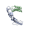 6z3hSC  6z3lC  6z3mC C: citing same article ( S: Starting model for refinement |
|---|---|
| Similar structure data |


-Links
 PDBj
PDBj


- Assembly
| Deposited unit | 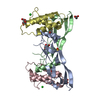
| ||||||||
|---|---|---|---|---|---|---|---|---|---|
| 1 |
| ||||||||
| Unit cell |
|
-Components
-Protein , 2 types, 4 molecules ABCD
| #1: Protein | Mass: 13358.446 Da / Num. of mol.: 2 / Mutation: Y487K, Q489D Source method: isolated from a genetically manipulated source Source: (gene. exp.) Homo sapiens (human) / Gene: GDF5, BMP14, CDMP1 / Production host:  Escherichia coli (E. coli) / References: UniProt: P43026 Escherichia coli (E. coli) / References: UniProt: P43026#2: Protein | Mass: 10532.701 Da / Num. of mol.: 2 Source method: isolated from a genetically manipulated source Source: (gene. exp.) Homo sapiens (human) / Gene: RGMB / Cell line (production host): HEK293T / Production host: Homo sapiens (human) / References: UniProt: Q6NW40 |
|---|
-Sugars , 1 types, 1 molecules 
| #7: Sugar | ChemComp-NAG / N-Acetylglucosamine Type: D-saccharide, beta linking / Mass: 221.208 Da / Num. of mol.: 1 Type: D-saccharide, beta linking / Mass: 221.208 Da / Num. of mol.: 1Source method: isolated from a genetically manipulated source Formula: C8H15NO6 / Feature type: SUBJECT OF INVESTIGATION |
|---|
-Non-polymers , 5 types, 152 molecules 


| #3: Chemical | Sulfate Mass: 96.063 Da / Num. of mol.: 2 / Source method: obtained synthetically / Formula: SO4 / Feature type: SUBJECT OF INVESTIGATION Mass: 96.063 Da / Num. of mol.: 2 / Source method: obtained synthetically / Formula: SO4 / Feature type: SUBJECT OF INVESTIGATION#4: Chemical | Glycerol Mass: 92.094 Da / Num. of mol.: 2 / Source method: obtained synthetically / Formula: C3H8O3 / Feature type: SUBJECT OF INVESTIGATION Mass: 92.094 Da / Num. of mol.: 2 / Source method: obtained synthetically / Formula: C3H8O3 / Feature type: SUBJECT OF INVESTIGATION#5: Chemical | Ethylene glycol Mass: 62.068 Da / Num. of mol.: 3 / Source method: obtained synthetically / Formula: C2H6O2 / Feature type: SUBJECT OF INVESTIGATION Mass: 62.068 Da / Num. of mol.: 3 / Source method: obtained synthetically / Formula: C2H6O2 / Feature type: SUBJECT OF INVESTIGATION#6: Chemical | Chloride Mass: 35.453 Da / Num. of mol.: 3 / Source method: obtained synthetically / Formula: Cl / Feature type: SUBJECT OF INVESTIGATION Mass: 35.453 Da / Num. of mol.: 3 / Source method: obtained synthetically / Formula: Cl / Feature type: SUBJECT OF INVESTIGATION#8: Water | ChemComp-HOH / | WaterMass: 18.015 Da / Num. of mol.: 142 / Source method: isolated from a natural source / Formula: H2O |
|---|
-Details
| Has ligand of interest | Y |
|---|
-Experimental details
-Experiment
| Experiment | Method: X-RAY DIFFRACTION / Number of used crystals: 1 |
|---|
- Sample preparation
| Crystal | Density Matthews: 1.92 Å3/Da / Density % sol: 35.96 % |
|---|---|
| Crystal grow | Temperature: 294 K / Method: vapor diffusion, sitting drop / pH: 7.4 Details: 0.2 M Li2SO4, 0.1 M HEPES pH 7.5, 25% v/v PEG 3350. |
-Data collection
| Diffraction | Mean temperature: 100 K / Serial crystal experiment: N |
|---|---|
| Diffraction source | Source: SYNCHROTRON / Site: Diamond / Beamline: I03 / Wavelength: 0.9762 Å |
| Detector | Type: DECTRIS PILATUS 6M / Detector: PIXEL / Date: Oct 25, 2017 |
| Radiation | Protocol: SINGLE WAVELENGTH / Monochromatic (M) / Laue (L): M / Scattering type: x-ray |
| Radiation wavelength | Wavelength: 0.9762 Å / Relative weight: 1 |
| Reflection | Resolution: 1.65→39.38 Å / Num. obs: 43071 / % possible obs: 99.7 % / Redundancy: 6.8 % / Rmerge(I) obs: 0.088 / Net I/σ(I): 9.5 |
| Reflection shell | Resolution: 1.65→1.69 Å / Mean I/σ(I) obs: 0.5 / Num. unique obs: 3117 / CC1/2: 0.212 |
- Processing
| Software |
| |||||||||||||||||||||||||||||||||||||||||||||||||||||||||||||||||||||||||||||||||||||||||||||||||||||||||||||||||||||||||||||
|---|---|---|---|---|---|---|---|---|---|---|---|---|---|---|---|---|---|---|---|---|---|---|---|---|---|---|---|---|---|---|---|---|---|---|---|---|---|---|---|---|---|---|---|---|---|---|---|---|---|---|---|---|---|---|---|---|---|---|---|---|---|---|---|---|---|---|---|---|---|---|---|---|---|---|---|---|---|---|---|---|---|---|---|---|---|---|---|---|---|---|---|---|---|---|---|---|---|---|---|---|---|---|---|---|---|---|---|---|---|---|---|---|---|---|---|---|---|---|---|---|---|---|---|---|---|---|
| Refinement | Method to determine structure: MOLECULAR REPLACEMENT Starting model: 6Z3H Resolution: 1.65→39.38 Å / SU ML: 0.3 / Cross valid method: FREE R-VALUE / σ(F): 1.33 / Phase error: 30.09 / Stereochemistry target values: ML
| |||||||||||||||||||||||||||||||||||||||||||||||||||||||||||||||||||||||||||||||||||||||||||||||||||||||||||||||||||||||||||||
| Solvent computation | Shrinkage radii: 0.9 Å / VDW probe radii: 1.11 Å / Solvent model: FLAT BULK SOLVENT MODEL | |||||||||||||||||||||||||||||||||||||||||||||||||||||||||||||||||||||||||||||||||||||||||||||||||||||||||||||||||||||||||||||
| Refinement step | Cycle: LAST / Resolution: 1.65→39.38 Å
| |||||||||||||||||||||||||||||||||||||||||||||||||||||||||||||||||||||||||||||||||||||||||||||||||||||||||||||||||||||||||||||
| Refine LS restraints |
| |||||||||||||||||||||||||||||||||||||||||||||||||||||||||||||||||||||||||||||||||||||||||||||||||||||||||||||||||||||||||||||
| LS refinement shell |
| |||||||||||||||||||||||||||||||||||||||||||||||||||||||||||||||||||||||||||||||||||||||||||||||||||||||||||||||||||||||||||||
| Refinement TLS params. | Method: refined / Refine-ID: X-RAY DIFFRACTION
| |||||||||||||||||||||||||||||||||||||||||||||||||||||||||||||||||||||||||||||||||||||||||||||||||||||||||||||||||||||||||||||
| Refinement TLS group |
|
