 Movie
Movie Controller

[English] 日本語
 Yorodumi
- PDB-6z3i: Structure of recombinant beta-glucocerebrosidase in complex with ... -
+ Open data

- Basic information
| Entry | Database: PDB / ID: 6z3i | ||||||
|---|---|---|---|---|---|---|---|
| Title | Structure of recombinant beta-glucocerebrosidase in complex with bifunctional cyclophellitol aziridine activity based probe | ||||||
 Components Components | Lysosomal acid glucosylceramidase | ||||||
 Keywords Keywords |  HYDROLASE / lysosomal hydrolase / activity based probe HYDROLASE / lysosomal hydrolase / activity based probe | ||||||
| Function / homology |  Function and homology information Function and homology informationpositive regulation of protein lipidation / steryl-beta-glucosidase activity / beta-glucoside catabolic process / positive regulation of neuronal action potential / cerebellar Purkinje cell layer formation / positive regulation of autophagy of mitochondrion in response to mitochondrial depolarization / galactosylceramidase / termination of signal transduction / galactosylceramidase activity / lymphocyte migration ...positive regulation of protein lipidation / steryl-beta-glucosidase activity / beta-glucoside catabolic process / positive regulation of neuronal action potential / cerebellar Purkinje cell layer formation / positive regulation of autophagy of mitochondrion in response to mitochondrial depolarization / galactosylceramidase / termination of signal transduction / galactosylceramidase activity / lymphocyte migration / glucosylceramidase / glucosylceramide catabolic process / scavenger receptor binding / regulation of lysosomal protein catabolic process / autophagosome organization / sphingosine biosynthetic process / glucosylceramidase activity / microglial cell proliferation / glucosyltransferase activity / regulation of TOR signaling / ceramide biosynthetic process / lipid storage / response to thyroid hormone / microglia differentiation / Glycosphingolipid catabolism / pyramidal neuron differentiation / lipid glycosylation / brain morphogenesis / Hydrolases; Glycosylases; Glycosidases, i.e. enzymes that hydrolyse O- and S-glycosyl compounds / response to pH / positive regulation of protein-containing complex disassembly / motor behavior / neuromuscular process / Transferases; Glycosyltransferases; Hexosyltransferases / hematopoietic stem cell proliferation / lysosome organization / response to testosterone / response to dexamethasone / Association of TriC/CCT with target proteins during biosynthesis / negative regulation of interleukin-6 production / antigen processing and presentation / homeostasis of number of cells / regulation of macroautophagy / establishment of skin barrier / negative regulation of protein-containing complex assembly / positive regulation of protein dephosphorylation / cell maturation / respiratory electron transport chain / cellular response to starvation / cholesterol metabolic process / lysosomal lumen / negative regulation of MAP kinase activity / determination of adult lifespan / trans-Golgi network / autophagy / negative regulation of inflammatory response / response to estrogen / positive regulation of proteasomal ubiquitin-dependent protein catabolic process / cellular response to tumor necrosis factor / T cell differentiation in thymus / proteasome-mediated ubiquitin-dependent protein catabolic process / neuron apoptotic process / negative regulation of neuron apoptotic process / lysosome / lysosomal membrane / signaling receptor binding / Golgi apparatus / endoplasmic reticulum / extracellular exosomeSimilarity search - Function | ||||||
| Biological species |  Homo sapiens (human) Homo sapiens (human) | ||||||
| Method | X-RAY DIFFRACTION / SYNCHROTRON / MOLECULAR REPLACEMENT / Resolution: 1.8 Å | ||||||
 Authors Authors | Rowland, R.J. / Davies, G.J. | ||||||
| Funding support |  United Kingdom, 1items United Kingdom, 1items
| ||||||
 Citation Citation | Journal: Chemistry / Year: 2021 Title: Design, Synthesis and Structural Analysis of Glucocerebrosidase Imaging Agents. Authors: Rowland, R.J. / Chen, Y. / Breen, I. / Wu, L. / Offen, W.A. / Beenakker, T.J. / Su, Q. / van den Nieuwendijk, A.M.C.H. / Aerts, J.M.F.G. / Artola, M. / Overkleeft, H.S. / Davies, G.J. | ||||||
| History |
|
- Structure visualization
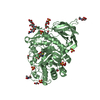
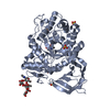
| Structure viewer | Molecule: MolmilJmol/JSmol |
|---|
- Downloads & links
-Download
| PDBx/mmCIF format | 6z3i.cif.gz | 222.1 KB | Display | PDBx/mmCIF format |
|---|---|---|---|---|
| PDB format | pdb6z3i.ent.gz | Display | PDB format | |
| PDBx/mmJSON format | 6z3i.json.gz | Tree view | PDBx/mmJSON format | |
| Others |  Other downloads Other downloads |
-Validation report
| Arichive directory | https://data.pdbj.org/pub/pdb/validation_reports/z3/6z3iftp://data.pdbj.org/pub/pdb/validation_reports/z3/6z3i | HTTPS FTP |
|---|
-Related structure data
| Related structure data | 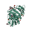 6ytpC 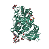 6ytrC 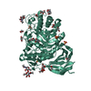 6yutC 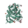 6yv3C 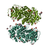 6z39C 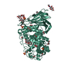 6tjkS C: citing same article ( S: Starting model for refinement |
|---|---|
| Similar structure data |


-Links
 PDBj
PDBj


- Assembly
| Deposited unit | 
| ||||||||
|---|---|---|---|---|---|---|---|---|---|
| 1 |
| ||||||||
| Unit cell |
|
-Components
-Protein , 1 types, 1 molecules BBB
| #1: Protein | Mass: 55659.219 Da / Num. of mol.: 1 Source method: isolated from a genetically manipulated source Details: Recombinant human beta-glucocerebrosidase lacking its 40 amino acid signalling sequence produced in insect cells Source: (gene. exp.) Homo sapiens (human) / Gene: GBA, GC, GLUC / Production host:  Trichoplusia ni (cabbage looper) Trichoplusia ni (cabbage looper)References: UniProt: P04062, glucosylceramidase, Transferases; Glycosyltransferases; Hexosyltransferases, steryl-beta-glucosidase |
|---|
-Sugars , 2 types, 3 molecules 
| #2: Polysaccharide | alpha-D-mannopyranose-(1-6)-beta-D-mannopyranose-(1-4)-2-acetamido-2-deoxy-beta-D-glucopyranose-(1- ...alpha-D-mannopyranose-(1-6)-beta-D-mannopyranose-(1-4)-2-acetamido-2-deoxy-beta-D-glucopyranose-(1-4)-2-acetamido-2-deoxy-beta-D-glucopyranose / Mass: 748.682 Da / Num. of mol.: 1 Source method: isolated from a genetically manipulated source |
|---|---|
| #4: Sugar | N-Acetylglucosamine Type: D-saccharide, beta linking / Mass: 221.208 Da / Num. of mol.: 2 Type: D-saccharide, beta linking / Mass: 221.208 Da / Num. of mol.: 2Source method: isolated from a genetically manipulated source Formula: C8H15NO6 |
-Non-polymers , 4 types, 359 molecules 


| #3: Chemical | ChemComp-SO4 / Sulfate Mass: 96.063 Da / Num. of mol.: 4 / Source method: obtained synthetically / Formula: SO4 Mass: 96.063 Da / Num. of mol.: 4 / Source method: obtained synthetically / Formula: SO4#5: Chemical | ChemComp-EDO / Ethylene glycol Mass: 62.068 Da / Num. of mol.: 16 Mass: 62.068 Da / Num. of mol.: 16Source method: isolated from a genetically manipulated source Formula: C2H6O2 #6: Chemical | ChemComp-Q68 / ~{ |  Mass: 383.486 Da / Num. of mol.: 1 Mass: 383.486 Da / Num. of mol.: 1Source method: isolated from a genetically manipulated source Formula: C18H33N5O4 / Feature type: SUBJECT OF INVESTIGATION #7: Water | ChemComp-HOH / | WaterMass: 18.015 Da / Num. of mol.: 338 / Source method: isolated from a natural source / Formula: H2O |
|---|
-Details
| Has ligand of interest | Y |
|---|
-Experimental details
-Experiment
| Experiment | Method: X-RAY DIFFRACTION / Number of used crystals: 1 |
|---|
- Sample preparation
| Crystal | Density Matthews: 2.43 Å3/Da / Density % sol: 49.45 % |
|---|---|
| Crystal grow | Temperature: 291 K / Method: vapor diffusion, sitting drop Details: 0.2 M Na2SO4, 0.25 M HEPES pH 7, PEG3350 14%, 1:1000 seed |
-Data collection
| Diffraction | Mean temperature: 100 K / Serial crystal experiment: N |
|---|---|
| Diffraction source | Source: SYNCHROTRON / Site: Diamond / Beamline: I04 / Wavelength: 0.979507 Å |
| Detector | Type: DECTRIS EIGER2 XE 16M / Detector: PIXEL / Date: Oct 19, 2019 |
| Radiation | Protocol: SINGLE WAVELENGTH / Monochromatic (M) / Laue (L): M / Scattering type: x-ray |
| Radiation wavelength | Wavelength: 0.979507 Å / Relative weight: 1 |
| Reflection | Resolution: 1.8→76.67 Å / Num. obs: 49541 / % possible obs: 100 % / Redundancy: 7 % / CC1/2: 0.995 / Rmerge(I) obs: 0.207 / Rpim(I) all: 0.084 / Net I/σ(I): 6.7 |
| Reflection shell | Resolution: 1.8→1.84 Å / Redundancy: 6.8 % / Rmerge(I) obs: 2.351 / Mean I/σ(I) obs: 0.8 / Num. unique obs: 2966 / CC1/2: 0.506 / Rpim(I) all: 1.301 / % possible all: 100 |
- Processing
| Software |
| |||||||||||||||||||||||||||||||||||||||||||||||||||||||||||||||||||||||||||||||||||||||||||||||||||||||||||||||||||||||||||||||||||||||||||||||||||||||||||
|---|---|---|---|---|---|---|---|---|---|---|---|---|---|---|---|---|---|---|---|---|---|---|---|---|---|---|---|---|---|---|---|---|---|---|---|---|---|---|---|---|---|---|---|---|---|---|---|---|---|---|---|---|---|---|---|---|---|---|---|---|---|---|---|---|---|---|---|---|---|---|---|---|---|---|---|---|---|---|---|---|---|---|---|---|---|---|---|---|---|---|---|---|---|---|---|---|---|---|---|---|---|---|---|---|---|---|---|---|---|---|---|---|---|---|---|---|---|---|---|---|---|---|---|---|---|---|---|---|---|---|---|---|---|---|---|---|---|---|---|---|---|---|---|---|---|---|---|---|---|---|---|---|---|---|---|---|
| Refinement | Method to determine structure: MOLECULAR REPLACEMENT Starting model: 6TJK Resolution: 1.8→66.578 Å / Cor.coef. Fo:Fc: 0.967 / Cor.coef. Fo:Fc free: 0.962 / WRfactor Rfree: 0.171 / WRfactor Rwork: 0.163 / SU B: 3.598 / SU ML: 0.098 / Average fsc free: 0.8785 / Average fsc work: 0.8756 / Cross valid method: FREE R-VALUE / ESU R: 0.129 / ESU R Free: 0.107 Details: Hydrogens have been added in their riding positions
| |||||||||||||||||||||||||||||||||||||||||||||||||||||||||||||||||||||||||||||||||||||||||||||||||||||||||||||||||||||||||||||||||||||||||||||||||||||||||||
| Solvent computation | Ion probe radii: 0.8 Å / Shrinkage radii: 0.8 Å / VDW probe radii: 1.2 Å / Solvent model: MASK BULK SOLVENT | |||||||||||||||||||||||||||||||||||||||||||||||||||||||||||||||||||||||||||||||||||||||||||||||||||||||||||||||||||||||||||||||||||||||||||||||||||||||||||
| Displacement parameters | Biso mean: 25.448 Å2
| |||||||||||||||||||||||||||||||||||||||||||||||||||||||||||||||||||||||||||||||||||||||||||||||||||||||||||||||||||||||||||||||||||||||||||||||||||||||||||
| Refinement step | Cycle: LAST / Resolution: 1.8→66.578 Å
| |||||||||||||||||||||||||||||||||||||||||||||||||||||||||||||||||||||||||||||||||||||||||||||||||||||||||||||||||||||||||||||||||||||||||||||||||||||||||||
| Refine LS restraints |
| |||||||||||||||||||||||||||||||||||||||||||||||||||||||||||||||||||||||||||||||||||||||||||||||||||||||||||||||||||||||||||||||||||||||||||||||||||||||||||
| LS refinement shell |
|
