 Movie
Movie Controller

[English] 日本語
 Yorodumi
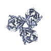
- PDB-6z0x: HtrA1 inactive protease domain S328A with CARASIL mutations D174R... -
+ Open data

- Basic information
| Entry | Database: PDB / ID: 6z0x | ||||||
|---|---|---|---|---|---|---|---|
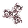
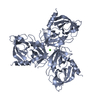
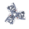
| Title | HtrA1 inactive protease domain S328A with CARASIL mutations D174R R274Q | ||||||
 Components Components | Serine protease HTRA1 | ||||||
 Keywords Keywords |  HYDROLASE / Hydrolase Protease HtrA family CARASIL mutations trimerization HYDROLASE / Hydrolase Protease HtrA family CARASIL mutations trimerization | ||||||
| Function / homology |  Function and homology information Function and homology informationchorionic trophoblast cell differentiation / programmed cell death / growth factor binding / negative regulation of BMP signaling pathway / Hydrolases; Acting on peptide bonds (peptidases); Serine endopeptidases / Degradation of the extracellular matrix / serine-type peptidase activity / placenta development / molecular function activator activity / negative regulation of transforming growth factor beta receptor signaling pathway ...chorionic trophoblast cell differentiation / programmed cell death / growth factor binding / negative regulation of BMP signaling pathway / Hydrolases; Acting on peptide bonds (peptidases); Serine endopeptidases / Degradation of the extracellular matrix / serine-type peptidase activity / placenta development / molecular function activator activity / negative regulation of transforming growth factor beta receptor signaling pathway / collagen-containing extracellular matrix / positive regulation of apoptotic process / serine-type endopeptidase activity / proteolysis / extracellular space / extracellular exosome / extracellular region / identical protein binding / plasma membrane / cytosolSimilarity search - Function | ||||||
| Biological species |  Homo sapiens (human) Homo sapiens (human) | ||||||
| Method | X-RAY DIFFRACTION / SYNCHROTRON / MOLECULAR REPLACEMENT / Resolution: 3.1 Å | ||||||
 Authors Authors | Vetter, I.R. / Stege, P. / Ingendahl, L. / Ehrmann, M. | ||||||
| Funding support |  Germany, 1items Germany, 1items
| ||||||
 Citation Citation | Journal: To Be Published Title: Repair strategies addressing pathogenic protein conformations Authors: Ingendahl, L. / Beaufort, N. / Kuszner, M. / Vetter, I.R. / Stege, P. / Ruiz-Blanco, Y.B. / Bravo-Rodriguez, K. / Beuck, C. / Schillinger, J. / Rey, J. / Roberti, A. / Hagemeier, B. / Hu, X.- ...Authors: Ingendahl, L. / Beaufort, N. / Kuszner, M. / Vetter, I.R. / Stege, P. / Ruiz-Blanco, Y.B. / Bravo-Rodriguez, K. / Beuck, C. / Schillinger, J. / Rey, J. / Roberti, A. / Hagemeier, B. / Hu, X.-Y. / Clausen, T. / Sanchez-Garcia, E. / Schmuck, C. / Dichgans, M. / Ehrmann, M. | ||||||
| History |
|
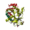
- Structure visualization
| Structure viewer | Molecule: MolmilJmol/JSmol |
|---|
- Downloads & links
-Download
| PDBx/mmCIF format | 6z0x.cif.gz | 130.3 KB | Display | PDBx/mmCIF format |
|---|---|---|---|---|
| PDB format | pdb6z0x.ent.gz | 101.2 KB | Display | PDB format |
| PDBx/mmJSON format | 6z0x.json.gz | Tree view | PDBx/mmJSON format | |
| Others |  Other downloads Other downloads |
-Validation report
| Arichive directory | https://data.pdbj.org/pub/pdb/validation_reports/z0/6z0xftp://data.pdbj.org/pub/pdb/validation_reports/z0/6z0x | HTTPS FTP |
|---|
-Related structure data
| Related structure data |  6z0eC  6z0yC  3tjoS C: citing same article ( S: Starting model for refinement |
|---|---|
| Similar structure data |

-Links
 PDBj
PDBj


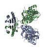
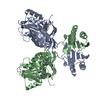
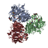
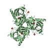
- Assembly
| Deposited unit | 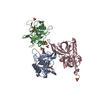
| ||||||||||||
|---|---|---|---|---|---|---|---|---|---|---|---|---|---|
| 1 | 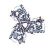
| ||||||||||||
| 2 | 
| ||||||||||||
| 3 | 
| ||||||||||||
| Unit cell |
| ||||||||||||
| Components on special symmetry positions |
|
-Components
| #1: Protein | Mass: 25659.359 Da / Num. of mol.: 3 / Mutation: D174R R274Q S328A Source method: isolated from a genetically manipulated source Source: (gene. exp.) Homo sapiens (human) / Gene: HTRA1, HTRA, PRSS11 / Production host:  Escherichia coli (E. coli) Escherichia coli (E. coli)References: UniProt: Q92743, Hydrolases; Acting on peptide bonds (peptidases); Serine endopeptidases#2: Chemical | ChemComp-SO4 / Sulfate  Mass: 96.063 Da / Num. of mol.: 9 / Source method: obtained synthetically / Formula: SO4 Mass: 96.063 Da / Num. of mol.: 9 / Source method: obtained synthetically / Formula: SO4#3: Water | ChemComp-HOH / | Water Mass: 18.015 Da / Num. of mol.: 14 / Source method: isolated from a natural source / Formula: H2O Mass: 18.015 Da / Num. of mol.: 14 / Source method: isolated from a natural source / Formula: H2OHas ligand of interest | N | |
|---|
-Experimental details
-Experiment
| Experiment | Method: X-RAY DIFFRACTION / Number of used crystals: 1 |
|---|
- Sample preparation
| Crystal | Density Matthews: 2.96 Å3/Da / Density % sol: 58.45 % |
|---|---|
| Crystal grow | Temperature: 293 K / Method: vapor diffusion, sitting drop / pH: 4 / Details: 1.6 M ammonium sulfate, 100 mM Citrate pH 4 |
-Data collection
| Diffraction | Mean temperature: 100 K / Serial crystal experiment: N | ||||||||||||||||||||||||||||||||||||||||||||||||||||||||||||||||||||||||||||||||||||||||||||||||||||||||||||||||||||||||||||||||||||||||||||||||||||||||||||||||||||||||||||||||||||||||||||||||||||||||||||||||||
|---|---|---|---|---|---|---|---|---|---|---|---|---|---|---|---|---|---|---|---|---|---|---|---|---|---|---|---|---|---|---|---|---|---|---|---|---|---|---|---|---|---|---|---|---|---|---|---|---|---|---|---|---|---|---|---|---|---|---|---|---|---|---|---|---|---|---|---|---|---|---|---|---|---|---|---|---|---|---|---|---|---|---|---|---|---|---|---|---|---|---|---|---|---|---|---|---|---|---|---|---|---|---|---|---|---|---|---|---|---|---|---|---|---|---|---|---|---|---|---|---|---|---|---|---|---|---|---|---|---|---|---|---|---|---|---|---|---|---|---|---|---|---|---|---|---|---|---|---|---|---|---|---|---|---|---|---|---|---|---|---|---|---|---|---|---|---|---|---|---|---|---|---|---|---|---|---|---|---|---|---|---|---|---|---|---|---|---|---|---|---|---|---|---|---|---|---|---|---|---|---|---|---|---|---|---|---|---|---|---|---|---|
| Diffraction source | Source: SYNCHROTRON / Site: SLS  / Beamline: X10SA / Wavelength: 0.91504 Å / Beamline: X10SA / Wavelength: 0.91504 Å | ||||||||||||||||||||||||||||||||||||||||||||||||||||||||||||||||||||||||||||||||||||||||||||||||||||||||||||||||||||||||||||||||||||||||||||||||||||||||||||||||||||||||||||||||||||||||||||||||||||||||||||||||||
| Detector | Type: DECTRIS PILATUS 6M-F / Detector: PIXEL / Date: Sep 24, 2018 | ||||||||||||||||||||||||||||||||||||||||||||||||||||||||||||||||||||||||||||||||||||||||||||||||||||||||||||||||||||||||||||||||||||||||||||||||||||||||||||||||||||||||||||||||||||||||||||||||||||||||||||||||||
| Radiation | Protocol: SINGLE WAVELENGTH / Monochromatic (M) / Laue (L): M / Scattering type: x-ray | ||||||||||||||||||||||||||||||||||||||||||||||||||||||||||||||||||||||||||||||||||||||||||||||||||||||||||||||||||||||||||||||||||||||||||||||||||||||||||||||||||||||||||||||||||||||||||||||||||||||||||||||||||
| Radiation wavelength | Wavelength: 0.91504 Å / Relative weight: 1 | ||||||||||||||||||||||||||||||||||||||||||||||||||||||||||||||||||||||||||||||||||||||||||||||||||||||||||||||||||||||||||||||||||||||||||||||||||||||||||||||||||||||||||||||||||||||||||||||||||||||||||||||||||
| Reflection | Resolution: 3.1→48.88 Å / Num. obs: 15480 / % possible obs: 95.4 % / Redundancy: 20.537 % / Biso Wilson estimate: 110.027 Å2 / CC1/2: 1 / Rmerge(I) obs: 0.114 / Rrim(I) all: 0.117 / Χ2: 1.012 / Net I/σ(I): 17.4 / Num. measured all: 317918 | ||||||||||||||||||||||||||||||||||||||||||||||||||||||||||||||||||||||||||||||||||||||||||||||||||||||||||||||||||||||||||||||||||||||||||||||||||||||||||||||||||||||||||||||||||||||||||||||||||||||||||||||||||
| Reflection shell | Diffraction-ID: 1
|
- Processing
| Software |
| |||||||||||||||||||||||||||||||||||||||||||||||||||||||||||||||||||||||||||
|---|---|---|---|---|---|---|---|---|---|---|---|---|---|---|---|---|---|---|---|---|---|---|---|---|---|---|---|---|---|---|---|---|---|---|---|---|---|---|---|---|---|---|---|---|---|---|---|---|---|---|---|---|---|---|---|---|---|---|---|---|---|---|---|---|---|---|---|---|---|---|---|---|---|---|---|---|
| Refinement | Method to determine structure: MOLECULAR REPLACEMENT Starting model: 3TJO Resolution: 3.1→48.88 Å / Cor.coef. Fo:Fc: 0.939 / Cor.coef. Fo:Fc free: 0.897 / SU B: 25.343 / SU ML: 0.415 / Cross valid method: THROUGHOUT / σ(F): 0 / ESU R Free: 0.475 Details: HYDROGENS HAVE BEEN ADDED IN THE RIDING POSITIONS U VALUES : REFINED INDIVIDUALLY
| |||||||||||||||||||||||||||||||||||||||||||||||||||||||||||||||||||||||||||
| Solvent computation | Ion probe radii: 0.8 Å / Shrinkage radii: 0.8 Å / VDW probe radii: 1.2 Å | |||||||||||||||||||||||||||||||||||||||||||||||||||||||||||||||||||||||||||
| Displacement parameters | Biso max: 233.06 Å2 / Biso mean: 132.071 Å2 / Biso min: 82.53 Å2
| |||||||||||||||||||||||||||||||||||||||||||||||||||||||||||||||||||||||||||
| Refinement step | Cycle: final / Resolution: 3.1→48.88 Å
| |||||||||||||||||||||||||||||||||||||||||||||||||||||||||||||||||||||||||||
| Refine LS restraints |
| |||||||||||||||||||||||||||||||||||||||||||||||||||||||||||||||||||||||||||
| LS refinement shell | Resolution: 3.1→3.18 Å / Rfactor Rfree error: 0 / Total num. of bins used: 20
|
