 Movie
Movie Controller

[English] 日本語
 Yorodumi
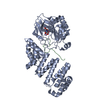
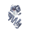
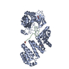
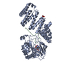
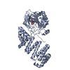
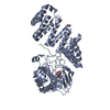
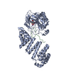
- PDB-6yyw: Aspartyl/Asparaginyl beta-hydroxylase (AspH) oxygenase and TPR do... -
+ Open data

- Basic information
| Entry | Database: PDB / ID: 6yyw | ||||||
|---|---|---|---|---|---|---|---|
| Title | Aspartyl/Asparaginyl beta-hydroxylase (AspH) oxygenase and TPR domains in complex with manganese, 2-oxoglutarate, and factor X substrate peptide fragment(39mer-4Ser) | ||||||
 Components Components |
| ||||||
 Keywords Keywords |  OXIDOREDUCTASE / Aspartyl/asparaginyl beta-hydroxylase / Dioxygenase OXIDOREDUCTASE / Aspartyl/asparaginyl beta-hydroxylase / Dioxygenase | ||||||
| Function / homology |  Function and homology informationpeptide-aspartate beta-dioxygenase / regulation of inositol 1,4,5-trisphosphate-sensitive calcium-release channel activity / regulation of protein depolymerization / activation of store-operated calcium channel activity / regulation of cell communication by electrical coupling / junctional sarcoplasmic reticulum membrane / peptidyl-aspartic acid 3-dioxygenase activity / cortical endoplasmic reticulum / sarcoplasmic reticulum lumen / limb morphogenesis ...peptide-aspartate beta-dioxygenase / regulation of inositol 1,4,5-trisphosphate-sensitive calcium-release channel activity / regulation of protein depolymerization / activation of store-operated calcium channel activity / regulation of cell communication by electrical coupling / junctional sarcoplasmic reticulum membrane / peptidyl-aspartic acid 3-dioxygenase activity / cortical endoplasmic reticulum / sarcoplasmic reticulum lumen / limb morphogenesis / coagulation factor Xa / pattern specification process / positive regulation of intracellular protein transport / Defective factor IX causes thrombophilia / Defective cofactor function of FVIIIa variant / Defective F9 variant does not activate FX / activation of cysteine-type endopeptidase activity / face morphogenesis / Extrinsic Pathway of Fibrin Clot Formation / structural constituent of muscle / positive regulation of calcium ion transport into cytosol / response to ATP / roof of mouth development / positive regulation of ryanodine-sensitive calcium-release channel activity / Protein hydroxylation / positive regulation of proteolysis / detection of calcium ion / positive regulation of TOR signaling / calcium ion homeostasis / regulation of cardiac muscle contraction by regulation of the release of sequestered calcium ion / Gamma-carboxylation of protein precursors / Transport of gamma-carboxylated protein precursors from the endoplasmic reticulum to the Golgi apparatus / Common Pathway of Fibrin Clot Formation / Removal of aminoterminal propeptides from gamma-carboxylated proteins / regulation of release of sequestered calcium ion into cytosol by sarcoplasmic reticulum / Ion homeostasis / regulation of ryanodine-sensitive calcium-release channel activity / sarcoplasmic reticulum membrane / calcium channel complex / cellular response to calcium ion / Intrinsic Pathway of Fibrin Clot Formation / muscle contraction / calcium ion transmembrane transport / phospholipid binding / regulation of protein stability / Stimuli-sensing channels / Golgi lumen / blood coagulation / cell population proliferation / transmembrane transporter binding / electron transfer activity / positive regulation of cell migration / negative regulation of cell population proliferation / external side of plasma membrane / endoplasmic reticulum lumen / serine-type endopeptidase activity / calcium ion binding / endoplasmic reticulum membrane / structural molecule activity / positive regulation of DNA-templated transcription / endoplasmic reticulum / proteolysis / extracellular space / extracellular region / plasma membrane Function and homology informationpeptide-aspartate beta-dioxygenase / regulation of inositol 1,4,5-trisphosphate-sensitive calcium-release channel activity / regulation of protein depolymerization / activation of store-operated calcium channel activity / regulation of cell communication by electrical coupling / junctional sarcoplasmic reticulum membrane / peptidyl-aspartic acid 3-dioxygenase activity / cortical endoplasmic reticulum / sarcoplasmic reticulum lumen / limb morphogenesis ...peptide-aspartate beta-dioxygenase / regulation of inositol 1,4,5-trisphosphate-sensitive calcium-release channel activity / regulation of protein depolymerization / activation of store-operated calcium channel activity / regulation of cell communication by electrical coupling / junctional sarcoplasmic reticulum membrane / peptidyl-aspartic acid 3-dioxygenase activity / cortical endoplasmic reticulum / sarcoplasmic reticulum lumen / limb morphogenesis / coagulation factor Xa / pattern specification process / positive regulation of intracellular protein transport / Defective factor IX causes thrombophilia / Defective cofactor function of FVIIIa variant / Defective F9 variant does not activate FX / activation of cysteine-type endopeptidase activity / face morphogenesis / Extrinsic Pathway of Fibrin Clot Formation / structural constituent of muscle / positive regulation of calcium ion transport into cytosol / response to ATP / roof of mouth development / positive regulation of ryanodine-sensitive calcium-release channel activity / Protein hydroxylation / positive regulation of proteolysis / detection of calcium ion / positive regulation of TOR signaling / calcium ion homeostasis / regulation of cardiac muscle contraction by regulation of the release of sequestered calcium ion / Gamma-carboxylation of protein precursors / Transport of gamma-carboxylated protein precursors from the endoplasmic reticulum to the Golgi apparatus / Common Pathway of Fibrin Clot Formation / Removal of aminoterminal propeptides from gamma-carboxylated proteins / regulation of release of sequestered calcium ion into cytosol by sarcoplasmic reticulum / Ion homeostasis / regulation of ryanodine-sensitive calcium-release channel activity / sarcoplasmic reticulum membrane / calcium channel complex / cellular response to calcium ion / Intrinsic Pathway of Fibrin Clot Formation / muscle contraction / calcium ion transmembrane transport / phospholipid binding / regulation of protein stability / Stimuli-sensing channels / Golgi lumen / blood coagulation / cell population proliferation / transmembrane transporter binding / electron transfer activity / positive regulation of cell migration / negative regulation of cell population proliferation / external side of plasma membrane / endoplasmic reticulum lumen / serine-type endopeptidase activity / calcium ion binding / endoplasmic reticulum membrane / structural molecule activity / positive regulation of DNA-templated transcription / endoplasmic reticulum / proteolysis / extracellular space / extracellular region / plasma membraneSimilarity search - Function | ||||||
| Biological species |  Homo sapiens (human) Homo sapiens (human) | ||||||
| Method | X-RAY DIFFRACTION / SYNCHROTRON / MOLECULAR REPLACEMENT / Resolution: 2.27 Å | ||||||
 Authors Authors | Nakashima, Y. / Brewitz, L. / Schofield, C.J. | ||||||
 Citation Citation | Journal: Chem Sci / Year: 2020 Title: Synthesis of 2-oxoglutarate derivatives and their evaluation as cosubstrates and inhibitors of human aspartate/asparagine-beta-hydroxylase. Authors: Brewitz, L. / Nakashima, Y. / Schofield, C.J. | ||||||
| History |
|
- Structure visualization
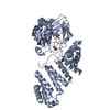
| Structure viewer | Molecule: MolmilJmol/JSmol |
|---|
- Downloads & links
-Download
| PDBx/mmCIF format | 6yyw.cif.gz | 210.3 KB | Display | PDBx/mmCIF format |
|---|---|---|---|---|
| PDB format | pdb6yyw.ent.gz | 144.2 KB | Display | PDB format |
| PDBx/mmJSON format | 6yyw.json.gz | Tree view | PDBx/mmJSON format | |
| Others |  Other downloads Other downloads |
-Validation report
| Arichive directory | https://data.pdbj.org/pub/pdb/validation_reports/yy/6yywftp://data.pdbj.org/pub/pdb/validation_reports/yy/6yyw | HTTPS FTP |
|---|
-Related structure data
| Related structure data |  6yyuC  6yyvC  6yyyC 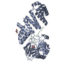 6z6qC  6z6rC  5jtcS S: Starting model for refinement C: citing same article ( |
|---|---|
| Similar structure data |


-Links
 PDBj
PDBj


- Assembly
| Deposited unit | 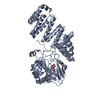
| ||||||||||||
|---|---|---|---|---|---|---|---|---|---|---|---|---|---|
| 1 |
| ||||||||||||
| Unit cell |
|
-Components
| #1: Protein | Mass: 49405.336 Da / Num. of mol.: 1 Source method: isolated from a genetically manipulated source Source: (gene. exp.) Homo sapiens (human) / Gene: ASPH, BAH / Production host:  Escherichia coli BL21(DE3) (bacteria) Escherichia coli BL21(DE3) (bacteria)References: UniProt: Q12797, peptide-aspartate beta-dioxygenase |
|---|---|
| #2: Protein/peptide | Factor X / Stuart factor / Stuart-Prower factor Mass: 4190.384 Da / Num. of mol.: 1 Source method: isolated from a genetically manipulated source Source: (gene. exp.) Homo sapiens (human) / Gene: F10 / Production host: synthetic construct (others) / References: UniProt: P00742, coagulation factor Xa |
| #3: Chemical | ChemComp-MN /   Mass: 54.938 Da / Num. of mol.: 1 / Source method: obtained synthetically / Formula: Mn / Feature type: SUBJECT OF INVESTIGATION Mass: 54.938 Da / Num. of mol.: 1 / Source method: obtained synthetically / Formula: Mn / Feature type: SUBJECT OF INVESTIGATION |
| #4: Chemical | ChemComp-AKG / Α-Ketoglutaric acid  Mass: 146.098 Da / Num. of mol.: 1 / Source method: obtained synthetically / Formula: C5H6O5 / Feature type: SUBJECT OF INVESTIGATION Mass: 146.098 Da / Num. of mol.: 1 / Source method: obtained synthetically / Formula: C5H6O5 / Feature type: SUBJECT OF INVESTIGATION |
| #5: Water | ChemComp-HOH / Water Mass: 18.015 Da / Num. of mol.: 143 / Source method: isolated from a natural source / Formula: H2O Mass: 18.015 Da / Num. of mol.: 143 / Source method: isolated from a natural source / Formula: H2O |
| Has ligand of interest | Y |
-Experimental details
-Experiment
| Experiment | Method: X-RAY DIFFRACTION / Number of used crystals: 1 |
|---|
- Sample preparation
| Crystal | Density Matthews: 2.75 Å3/Da / Density % sol: 55.33 % |
|---|---|
| Crystal grow | Temperature: 277 K / Method: vapor diffusion, sitting drop / pH: 7.5 Details: 100 mM Bis Tris Propane, 200 mM sodium acetate trihydrate, 20% w/v PEG 3350, 1 mM manganese chloride, 2 mM 2-oxoglutarate, 18 mg/ml protein |
-Data collection
| Diffraction | Mean temperature: 100 K / Serial crystal experiment: N |
|---|---|
| Diffraction source | Source: SYNCHROTRON / Site: Diamond  / Beamline: I04 / Wavelength: 0.97624 Å / Beamline: I04 / Wavelength: 0.97624 Å |
| Detector | Type: DECTRIS EIGER2 XE 16M / Detector: PIXEL / Date: Jan 19, 2020 |
| Radiation | Protocol: SINGLE WAVELENGTH / Monochromatic (M) / Laue (L): M / Scattering type: x-ray |
| Radiation wavelength | Wavelength: 0.97624 Å / Relative weight: 1 |
| Reflection | Resolution: 2.27→29.64 Å / Num. obs: 26364 / % possible obs: 97.2 % / Redundancy: 12.6 % / Biso Wilson estimate: 45 Å2 / CC1/2: 0.998 / Rmerge(I) obs: 0.125 / Net I/σ(I): 12 |
| Reflection shell | Resolution: 2.27→2.34 Å / Rmerge(I) obs: 0.863 / Mean I/σ(I) obs: 1.8 / Num. unique obs: 1700 / CC1/2: 0.807 / % possible all: 69.9 |
- Processing
| Software |
| |||||||||||||||||||||||||||||||||||||||||||||||||||||||||||||||||||||||||||||||||||||||||||||||||||||||||
|---|---|---|---|---|---|---|---|---|---|---|---|---|---|---|---|---|---|---|---|---|---|---|---|---|---|---|---|---|---|---|---|---|---|---|---|---|---|---|---|---|---|---|---|---|---|---|---|---|---|---|---|---|---|---|---|---|---|---|---|---|---|---|---|---|---|---|---|---|---|---|---|---|---|---|---|---|---|---|---|---|---|---|---|---|---|---|---|---|---|---|---|---|---|---|---|---|---|---|---|---|---|---|---|---|---|---|
| Refinement | Method to determine structure: MOLECULAR REPLACEMENT Starting model: 5JTC Resolution: 2.27→29.64 Å / SU ML: 0.286 / Cross valid method: FREE R-VALUE / σ(F): 1.35 / Phase error: 25.1051
| |||||||||||||||||||||||||||||||||||||||||||||||||||||||||||||||||||||||||||||||||||||||||||||||||||||||||
| Solvent computation | Shrinkage radii: 0.9 Å / VDW probe radii: 1.11 Å | |||||||||||||||||||||||||||||||||||||||||||||||||||||||||||||||||||||||||||||||||||||||||||||||||||||||||
| Displacement parameters | Biso mean: 56.89 Å2 | |||||||||||||||||||||||||||||||||||||||||||||||||||||||||||||||||||||||||||||||||||||||||||||||||||||||||
| Refinement step | Cycle: LAST / Resolution: 2.27→29.64 Å
| |||||||||||||||||||||||||||||||||||||||||||||||||||||||||||||||||||||||||||||||||||||||||||||||||||||||||
| Refine LS restraints |
| |||||||||||||||||||||||||||||||||||||||||||||||||||||||||||||||||||||||||||||||||||||||||||||||||||||||||
| LS refinement shell |
|
