 Movie
Movie Controller


+ Open data

- Basic information
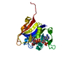
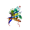
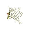
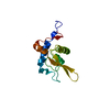
| Entry | Database: PDB / ID: 6yop | ||||||
|---|---|---|---|---|---|---|---|
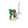
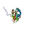
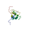
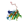
| Title | Structure of SAMM50 LIR bound to GABARAP | ||||||
 Components Components | Sorting and assembly machinery component 50 homolog,Gamma-aminobutyric acid receptor-associated protein | ||||||
 Keywords Keywords |  SIGNALING PROTEIN / LIR / SAMM50 / ATG8 SIGNALING PROTEIN / LIR / SAMM50 / ATG8 | ||||||
| Function / homology |  Function and homology information Function and homology informationMIB complex / SAM complex / inner mitochondrial membrane organization / Cristae formation / mitochondrial respiratory chain complex assembly / positive regulation of protein K48-linked ubiquitination / cristae formation / regulation of Rac protein signal transduction / GABA receptor binding / Mitochondrial protein import ...MIB complex / SAM complex / inner mitochondrial membrane organization / Cristae formation / mitochondrial respiratory chain complex assembly / positive regulation of protein K48-linked ubiquitination / cristae formation / regulation of Rac protein signal transduction / GABA receptor binding / Mitochondrial protein import / cellular response to nitrogen starvation / phosphatidylethanolamine binding / TBC/RABGAPs / microtubule associated complex / protein insertion into mitochondrial outer membrane / Macroautophagy / beta-tubulin binding / axoneme / autophagosome membrane / autophagosome assembly / extrinsic apoptotic signaling pathway via death domain receptors / smooth endoplasmic reticulum / autophagosome / RAC2 GTPase cycle / protein targeting / sperm midpiece / macroautophagy / microtubule cytoskeleton organization / positive regulation of proteasomal ubiquitin-dependent protein catabolic process / actin cytoskeleton / protein transport / cytoplasmic vesicle / microtubule binding / chemical synaptic transmission / microtubule / mitochondrial outer membrane / lysosome / Golgi membrane / synapse / ubiquitin protein ligase binding / mitochondrion / extracellular exosome / membrane / plasma membrane / cytosolSimilarity search - Function | ||||||
| Biological species |  Homo sapiens (human) Homo sapiens (human) | ||||||
| Method | X-RAY DIFFRACTION / SYNCHROTRON / MOLECULAR REPLACEMENT / Resolution: 1.1 Å | ||||||
 Authors Authors | Mouilleron, S. / Wenxin, Z. / Johansen, T. / Tooze, S. / Abudu, Y.P. / Lamark, T. | ||||||
 Citation Citation | Journal: To Be Published Title: Structure of SAMM50 LIR bound to GABARAP Authors: Mouilleron, S. / Wenxin, Z. / Johansen, T. / Tooze, S. / Abudu, Y.P. / Lamark, T. | ||||||
| History |
|
- Structure visualization
| Structure viewer | Molecule: MolmilJmol/JSmol |
|---|
- Downloads & links
-Download
| PDBx/mmCIF format | 6yop.cif.gz | 127.1 KB | Display | PDBx/mmCIF format |
|---|---|---|---|---|
| PDB format | pdb6yop.ent.gz | 85.4 KB | Display | PDB format |
| PDBx/mmJSON format | 6yop.json.gz | Tree view | PDBx/mmJSON format | |
| Others |  Other downloads Other downloads |
-Validation report
| Arichive directory | https://data.pdbj.org/pub/pdb/validation_reports/yo/6yopftp://data.pdbj.org/pub/pdb/validation_reports/yo/6yop | HTTPS FTP |
|---|
-Related structure data
| Related structure data |  1gnuS S: Starting model for refinement |
|---|---|
| Similar structure data |


-Links
 PDBj
PDBj


- Assembly
| Deposited unit | 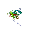
| ||||||||||||
|---|---|---|---|---|---|---|---|---|---|---|---|---|---|
| 1 |
| ||||||||||||
| Unit cell |
| ||||||||||||
| Components on special symmetry positions |
|
-Components
| #1: Protein | Mass: 15945.150 Da / Num. of mol.: 1 Source method: isolated from a genetically manipulated source Source: (gene. exp.) Homo sapiens (human) / Gene: SAMM50, SAM50, CGI-51, TRG3, GABARAP, FLC3B, HT004 / Production host:  Escherichia coli (E. coli) / References: UniProt: Q9Y512, UniProt: O95166 Escherichia coli (E. coli) / References: UniProt: Q9Y512, UniProt: O95166 |
|---|---|
| #2: Water | ChemComp-HOH / Water Mass: 18.015 Da / Num. of mol.: 177 / Source method: isolated from a natural source / Formula: H2O Mass: 18.015 Da / Num. of mol.: 177 / Source method: isolated from a natural source / Formula: H2O |
-Experimental details
-Experiment
| Experiment | Method: X-RAY DIFFRACTION / Number of used crystals: 1 |
|---|
- Sample preparation
| Crystal | Density Matthews: 2.76 Å3/Da / Density % sol: 55.36 % |
|---|---|
| Crystal grow | Temperature: 293 K / Method: vapor diffusion, sitting drop / Details: 20% v/v isopropanol, 0.1 M Na Acetate pH 5.5 |
-Data collection
| Diffraction | Mean temperature: 100 K / Serial crystal experiment: N |
|---|---|
| Diffraction source | Source: SYNCHROTRON / Site: Diamond  / Beamline: I04-1 / Wavelength: 0.97 Å / Beamline: I04-1 / Wavelength: 0.97 Å |
| Detector | Type: DECTRIS PILATUS 6M / Detector: PIXEL / Date: Jan 24, 2020 |
| Radiation | Protocol: SINGLE WAVELENGTH / Monochromatic (M) / Laue (L): M / Scattering type: x-ray |
| Radiation wavelength | Wavelength: 0.97 Å / Relative weight: 1 |
| Reflection | Resolution: 1.1→50.66 Å / Num. obs: 5665274 / % possible obs: 99.9 % / Redundancy: 81.1 % / Biso Wilson estimate: 15.19 Å2 / CC1/2: 1 / Rmerge(I) obs: 0.08 / Rpim(I) all: 0.01 / Net I/σ(I): 27.3 |
| Reflection shell | Resolution: 1.1→1.14 Å / Redundancy: 81.8 % / Rmerge(I) obs: 2.64 / Mean I/σ(I) obs: 0.9 / Num. unique obs: 567531 / CC1/2: 0.76 / Rpim(I) all: 0.29 / % possible all: 100 |
- Processing
| Software |
| ||||||||||||||||||||||||||||||||||||||||||||||||||||||||||||||||||||||||||||||||||||||||||||||||||||||||||||||||||||||||||||||||||||||||||||||||||||||||||||||||||||||||||||||||||||||
|---|---|---|---|---|---|---|---|---|---|---|---|---|---|---|---|---|---|---|---|---|---|---|---|---|---|---|---|---|---|---|---|---|---|---|---|---|---|---|---|---|---|---|---|---|---|---|---|---|---|---|---|---|---|---|---|---|---|---|---|---|---|---|---|---|---|---|---|---|---|---|---|---|---|---|---|---|---|---|---|---|---|---|---|---|---|---|---|---|---|---|---|---|---|---|---|---|---|---|---|---|---|---|---|---|---|---|---|---|---|---|---|---|---|---|---|---|---|---|---|---|---|---|---|---|---|---|---|---|---|---|---|---|---|---|---|---|---|---|---|---|---|---|---|---|---|---|---|---|---|---|---|---|---|---|---|---|---|---|---|---|---|---|---|---|---|---|---|---|---|---|---|---|---|---|---|---|---|---|---|---|---|---|---|
| Refinement | Method to determine structure: MOLECULAR REPLACEMENT Starting model: 1GNU Resolution: 1.1→50.66 Å / SU ML: 0.1054 / Cross valid method: FREE R-VALUE / σ(F): 1.35 / Phase error: 14.3818
| ||||||||||||||||||||||||||||||||||||||||||||||||||||||||||||||||||||||||||||||||||||||||||||||||||||||||||||||||||||||||||||||||||||||||||||||||||||||||||||||||||||||||||||||||||||||
| Solvent computation | Shrinkage radii: 0.9 Å / VDW probe radii: 1.11 Å | ||||||||||||||||||||||||||||||||||||||||||||||||||||||||||||||||||||||||||||||||||||||||||||||||||||||||||||||||||||||||||||||||||||||||||||||||||||||||||||||||||||||||||||||||||||||
| Displacement parameters | Biso mean: 21.96 Å2 | ||||||||||||||||||||||||||||||||||||||||||||||||||||||||||||||||||||||||||||||||||||||||||||||||||||||||||||||||||||||||||||||||||||||||||||||||||||||||||||||||||||||||||||||||||||||
| Refinement step | Cycle: LAST / Resolution: 1.1→50.66 Å
| ||||||||||||||||||||||||||||||||||||||||||||||||||||||||||||||||||||||||||||||||||||||||||||||||||||||||||||||||||||||||||||||||||||||||||||||||||||||||||||||||||||||||||||||||||||||
| Refine LS restraints |
| ||||||||||||||||||||||||||||||||||||||||||||||||||||||||||||||||||||||||||||||||||||||||||||||||||||||||||||||||||||||||||||||||||||||||||||||||||||||||||||||||||||||||||||||||||||||
| LS refinement shell |
|
