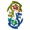
 Movie
Movie Controller


+ Open data

- Basic information
| Entry | Database: PDB / ID: 6ygm | ||||||
|---|---|---|---|---|---|---|---|
| Title | TGT W95F mutant labelled mit 5F-Trp crystallised at pH 5.5 | ||||||
 Components Components | Queuine tRNA-ribosyltransferase | ||||||
 Keywords Keywords | TRANSFERASE / TGT / TRNA-GUANINE TRANSGLYCOSYLASE / GUANINE INSERTION ENZYME / HOMODIMER / 19F-NMR / 5F-TRP | ||||||
| Function / homology |  Function and homology information Function and homology informationtRNA-guanosine34 preQ1 transglycosylase / tRNA wobble guanine modification / tRNA-guanosine(34) queuine transglycosylase activity / tRNA-guanine transglycosylation / queuosine biosynthetic process / metal ion binding / cytosolSimilarity search - Function | ||||||
| Biological species |  Zymomonas mobilis subsp. mobilis (bacteria) Zymomonas mobilis subsp. mobilis (bacteria) | ||||||
| Method | X-RAY DIFFRACTION / SYNCHROTRON / MOLECULAR REPLACEMENT / Resolution: 1.23 Å | ||||||
 Authors Authors | Nguyen, A. / Heine, A. / Klebe, G. | ||||||
 Citation Citation | Journal: To Be Published Title: Mutation study on tRNA-guanine transglycosylase within 19F NMR experiments for conformational change analysis Authors: Nguyen, A. / Heine, A. / Klebe, G. | ||||||
| History |
|
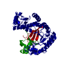
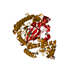
- Structure visualization
| Structure viewer | Molecule: MolmilJmol/JSmol |
|---|
- Downloads & links
-Download
| PDBx/mmCIF format | 6ygm.cif.gz | 267.6 KB | Display | PDBx/mmCIF format |
|---|---|---|---|---|
| PDB format | pdb6ygm.ent.gz | 180.4 KB | Display | PDB format |
| PDBx/mmJSON format | 6ygm.json.gz | Tree view | PDBx/mmJSON format | |
| Others |  Other downloads Other downloads |
-Validation report
| Arichive directory | https://data.pdbj.org/pub/pdb/validation_reports/yg/6ygmftp://data.pdbj.org/pub/pdb/validation_reports/yg/6ygm | HTTPS FTP |
|---|
-Related structure data
| Related structure data | 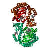 6yglC 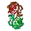 6ygoC 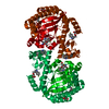 6ygpC  6ygwC 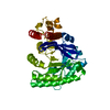 1pudS C: citing same article ( S: Starting model for refinement |
|---|---|
| Similar structure data |


-Links
 PDBj
PDBj- Assembly
| Deposited unit | 
| ||||||||||||
|---|---|---|---|---|---|---|---|---|---|---|---|---|---|
| 1 | 
| ||||||||||||
| Unit cell |
|
-Components
| #1: Protein | / Guanine insertion enzyme / tRNA-guanine transglycosylase Mass: 43084.766 Da / Num. of mol.: 1 / Mutation: T312K Source method: isolated from a genetically manipulated source Source: (gene. exp.) Zymomonas mobilis subsp. mobilis (strain ATCC 31821 / ZM4 / CP4) (bacteria)Gene: tgt, ZMO0363 / Production host: Escherichia coli (E. coli)References: UniProt: P28720, tRNA-guanosine34 preQ1 transglycosylase |
|---|---|
| #2: Chemical | ChemComp-ZN /   Mass: 65.409 Da / Num. of mol.: 1 / Source method: obtained synthetically / Formula: Zn Mass: 65.409 Da / Num. of mol.: 1 / Source method: obtained synthetically / Formula: Zn |
| #3: Water | ChemComp-HOH / Water Mass: 18.015 Da / Num. of mol.: 334 / Source method: isolated from a natural source / Formula: H2O Mass: 18.015 Da / Num. of mol.: 334 / Source method: isolated from a natural source / Formula: H2O |
| Has ligand of interest | N |
-Experimental details
-Experiment
| Experiment | Method: X-RAY DIFFRACTION / Number of used crystals: 1 |
|---|
- Sample preparation
| Crystal | Density Matthews: 2.27 Å3/Da / Density % sol: 45.7 % |
|---|---|
| Crystal grow | Temperature: 288 K / Method: vapor diffusion, hanging drop / pH: 5.5 Details: 100 mM MES pH 5.5, 0.5 mM DTT, 10% (v/v) DMSO, 13% (m/v) PEG8000 |
-Data collection
| Diffraction | Mean temperature: 100 K / Serial crystal experiment: N |
|---|---|
| Diffraction source | Source: SYNCHROTRON / Site: BESSY  / Beamline: 14.1 / Wavelength: 0.9184 Å / Beamline: 14.1 / Wavelength: 0.9184 Å |
| Detector | Type: DECTRIS PILATUS 6M / Detector: PIXEL / Date: Jul 11, 2019 |
| Radiation | Protocol: SINGLE WAVELENGTH / Monochromatic (M) / Laue (L): M / Scattering type: x-ray |
| Radiation wavelength | Wavelength: 0.9184 Å / Relative weight: 1 |
| Reflection | Resolution: 1.23→42.17 Å / Num. obs: 107425 / % possible obs: 97.5 % / Redundancy: 3.8 % / Biso Wilson estimate: 14.31 Å2 / CC1/2: 0.999 / Net I/σ(I): 16.11 |
| Reflection shell | Resolution: 1.23→1.31 Å / Num. unique obs: 16748 / CC1/2: 0.842 |
- Processing
| Software |
| |||||||||||||||||||||||||||||||||||||||||||||||||||||||||||||||||||||||||||||||||||||||||||||||||||||||||||||||||||||||||||||||||||||||||||||||||||||||||||||||||||||||||||||||||||||||||||||||||||||||||||||||||||||||||
|---|---|---|---|---|---|---|---|---|---|---|---|---|---|---|---|---|---|---|---|---|---|---|---|---|---|---|---|---|---|---|---|---|---|---|---|---|---|---|---|---|---|---|---|---|---|---|---|---|---|---|---|---|---|---|---|---|---|---|---|---|---|---|---|---|---|---|---|---|---|---|---|---|---|---|---|---|---|---|---|---|---|---|---|---|---|---|---|---|---|---|---|---|---|---|---|---|---|---|---|---|---|---|---|---|---|---|---|---|---|---|---|---|---|---|---|---|---|---|---|---|---|---|---|---|---|---|---|---|---|---|---|---|---|---|---|---|---|---|---|---|---|---|---|---|---|---|---|---|---|---|---|---|---|---|---|---|---|---|---|---|---|---|---|---|---|---|---|---|---|---|---|---|---|---|---|---|---|---|---|---|---|---|---|---|---|---|---|---|---|---|---|---|---|---|---|---|---|---|---|---|---|---|---|---|---|---|---|---|---|---|---|---|---|---|---|---|---|---|
| Refinement | Method to determine structure: MOLECULAR REPLACEMENT Starting model: 1PUD Resolution: 1.23→42.17 Å / SU ML: 0.1062 / Cross valid method: FREE R-VALUE / σ(F): 1.36 / Phase error: 15.8193
| |||||||||||||||||||||||||||||||||||||||||||||||||||||||||||||||||||||||||||||||||||||||||||||||||||||||||||||||||||||||||||||||||||||||||||||||||||||||||||||||||||||||||||||||||||||||||||||||||||||||||||||||||||||||||
| Solvent computation | Shrinkage radii: 0.9 Å / VDW probe radii: 1.11 Å | |||||||||||||||||||||||||||||||||||||||||||||||||||||||||||||||||||||||||||||||||||||||||||||||||||||||||||||||||||||||||||||||||||||||||||||||||||||||||||||||||||||||||||||||||||||||||||||||||||||||||||||||||||||||||
| Displacement parameters | Biso mean: 21.31 Å2 | |||||||||||||||||||||||||||||||||||||||||||||||||||||||||||||||||||||||||||||||||||||||||||||||||||||||||||||||||||||||||||||||||||||||||||||||||||||||||||||||||||||||||||||||||||||||||||||||||||||||||||||||||||||||||
| Refinement step | Cycle: LAST / Resolution: 1.23→42.17 Å
| |||||||||||||||||||||||||||||||||||||||||||||||||||||||||||||||||||||||||||||||||||||||||||||||||||||||||||||||||||||||||||||||||||||||||||||||||||||||||||||||||||||||||||||||||||||||||||||||||||||||||||||||||||||||||
| Refine LS restraints |
| |||||||||||||||||||||||||||||||||||||||||||||||||||||||||||||||||||||||||||||||||||||||||||||||||||||||||||||||||||||||||||||||||||||||||||||||||||||||||||||||||||||||||||||||||||||||||||||||||||||||||||||||||||||||||
| LS refinement shell |
|
